Clinical Trial
. 2020 Apr 3:11:529.
doi: 10.3389/fimmu.2020.00529. eCollection 2020.

CCR4+ Skin-Tropic Phenotype as a Feature of Central Memory CD8+ T Cells in Healthy Subjects and Psoriasis Patients

Affiliations
Clinical Trial

CCR4+ Skin-Tropic Phenotype as a Feature of Central Memory CD8+ T Cells in Healthy Subjects and Psoriasis Patients

Fabio Casciano et al. Front Immunol. .

Abstract

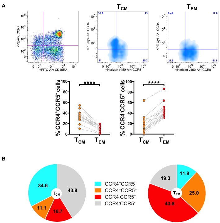
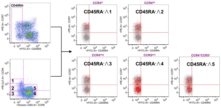
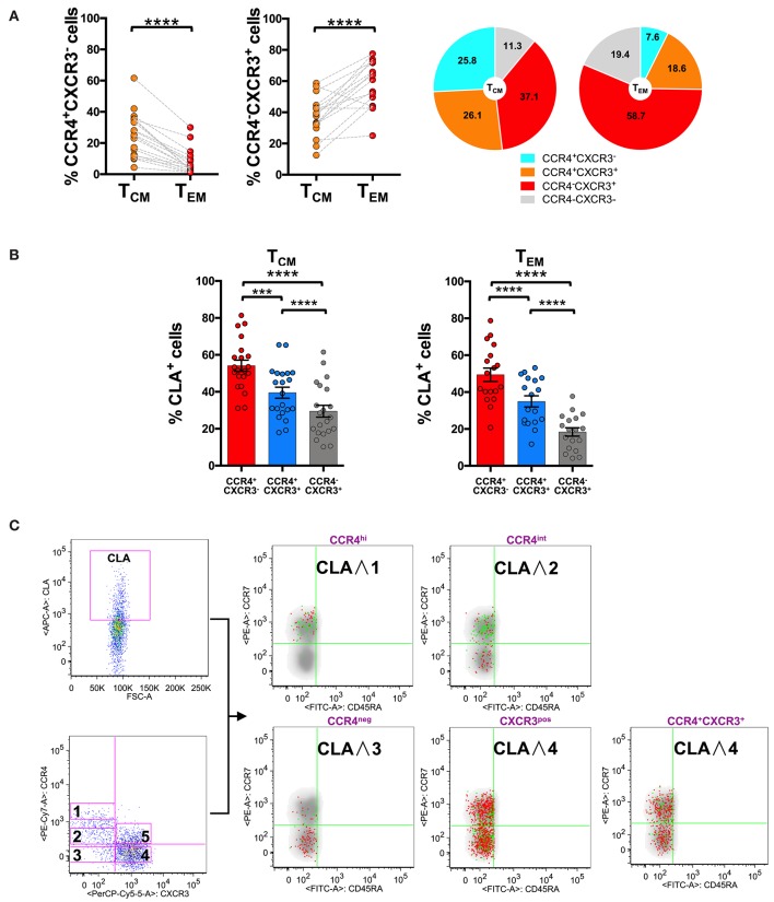
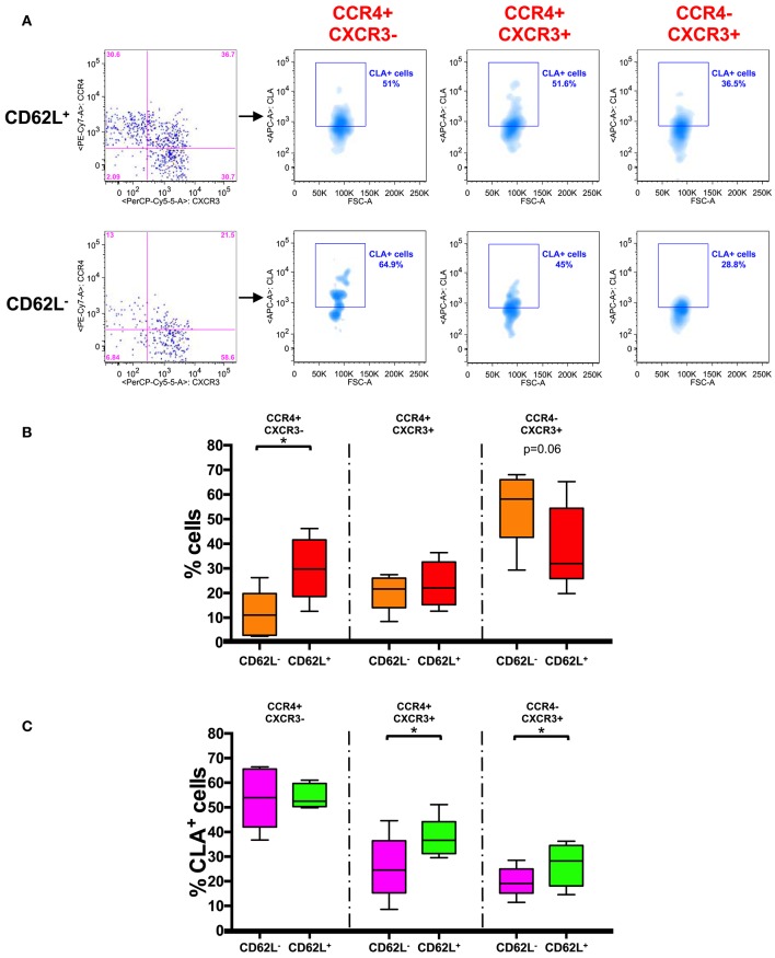
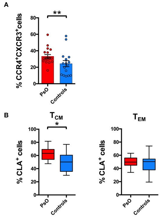
The chemokine receptor CCR4 has emerged as a skin-homing molecule important for the migration of T cells from the blood to the dermis. From our previous data on psoriasis patients, CCR4+ memory T cells emerged as a putative recirculating population between skin and blood. Here we focused our attention on the expression of CCR4 and skin-tropic molecules in the different stages of memory T cell differentiation. We analyzed the chemokine receptor profile in CD8+ and CD4+ CD45RA-CCR7+ (TCM) and CD45RA-CCR7- (TEM) cells. Subpopulations were further divided on the basis of CD62L expression, and the distribution among the subsets of the skin-homing molecule CLA (Cutaneous Lymphocyte Antigen) was evaluated. The characterization was performed on peripheral blood mononuclear cells isolated from 21 healthy subjects and 24 psoriasis patients. The results indicate that (i) the skin-homing CCR4 marker is mainly expressed in TCM cells, (ii) CCR4+ TCM cells also express high level of CLA and that (iii) the more differentiated phenotype TEM expresses CXCR3 and CCR5 but lower level of CCR4 and CLA. This indicates that progressive stages of memory T cell differentiation have profoundly different chemokine receptor patterns, with CD8+ TCM displaying a marked skin-tropic phenotype CLA+CCR4+. Differential skin-tropic phenotype between TCM and TEM cells was observed in both healthy subjects and psoriasis patients. However, patients showed an expanded circulating population of CD8+ TCM cells with phenotype CCR4+CXCR3+ that could play a role in the pathophysiology of psoriasis and possibly in disease recurrence.

Trial registration: ClinicalTrials.gov NCT03374527.

Keywords: T cells; central memory; effector memory; psoriatic disease; skin; tissue immunosurveillance.


Figures

Figure 1
Figure 1
Differential expression of CCR4 in CD8+ TCM and TEM cells. PBMCs isolated from healthy control subjects were stained for CD8, memory T cell phenotype markers (CD45RA and CCR7) and for chemokine receptors CCR4 and CCR5. (A) CD8+ T cells gated as CD45RACCR7+ TCM and CD45RACCR7 TEM were analyzed for the expression of CCR4 and CCR5. Representative analysis is shown in the figure. The axis scales for fluorescence are reported as log. Statistical analysis of the differences was performed by Mann–Whitney test. p-values < 0.05 were considered significant: ****p < 0.0001. (B) Mean values of the percentage of CCR4/CCR5 subpopulations among TCM and TEM cells were shown in pie charts.
Figure 2
Figure 2
CCR4 expression characterizes the TCM compartment. PBMCs isolated from healthy control subjects were stained for CD8, memory T cell phenotype markers (CD45RA and CCR7) and for chemokine receptors CCR4 and CCR5. (A) CD8+ T cells were analyzed for the memory phenotype according to CD45RA, CCR7 expression and for the expression of the chemokine receptors CCR4 and CCR5. On the basis of the chemokine receptor expression we identified five subsets CCR4hi (gate 1), CCR4int (gate 2), CCR4CCR5 (gate 3), CCR4CCR5+ (gate 4) and CCR4+CCR5+ (gate 5). These five subsets were superimposed to the density plot of the CD45RA gated cells. Each red dot identifies cells from the corresponding subset as reported in the figure. The axis scales for fluorescence are reported as log.
Figure 3
Figure 3
CLA expression is maximal in CCR4+ cells and considerably decreases in CXCR3+ cells. PBMCs isolated from healthy control subjects were stained for CD8, memory T cell phenotype markers (CD45RA and CCR7), for chemokine receptors CCR4, CXCR3, and for CLA. (A) CD8+ T cells gated as TCM and TEM were analyzed for the expression of CCR4 and CXCR3. Statistical analysis of the differences was performed by Mann-Whitney test. p-values < 0.05 were considered significant: ***p < 0.001, ****p < 0.0001. Mean values for CCR4/CXCR3 subpopulations among TCM and TEM cells were shown in pie charts. (B) TCM and TEM cells were analyzed for differences in the percentage of CLA+ cells in the CCR4+CXCR3, CCR4+CXCR3+, and CXCR3+ subpopulations using Mann-Whitney test. p-values < 0.05 were considered significant: ****p < 0.0001. (C) Reverse analysis is shown in the panel. Total memory CD45RA CD8+ T cells were gated and analyzed for the expression of CLA and for chemokine receptors CCR4/CXCR3. On the basis of the chemokine receptor expression we identified five subsets CCR4hi (gate 1), CCR4int (gate 2), CCR4CXCR3 (gate 3), CCR4CXCR3+ (gate 4), CCR4+CXCR3+ (gate 5). The five subsets of CCR4/CXCR3 expressing cells were overlaid to the density plot of CD45RA cells. Each red dot represents a cell from the corresponding subset. Among those dots, green dots identify cells of the gated subsets (CCR4hi, CCR4int, CCR4CXCR3, CCR4CXCR3+, and CCR4+CXCR3+) that co-express CLA. The axis scales for fluorescence are reported as log.
Figure 4
Figure 4
CD62L+ CCR7+ central memory T cells have a skin-tropic phenotype. (A) Representative analysis of chemokine receptor expression in CD62L+/− gated CD45RA CCR7+ (TCM) CD8+ T cells. Each subset (CCR4+CXCR3, CCR4+CXCR3+, and CCR4CXCR3+) in CD62L+ and CD62L gates was also analyzed for the expression of CLA. The axis scales for fluorescence are reported as log and the axis scale for FSC is reported as linear. (B) CD45RACCR7+ (TCM) CD8+ T cells were gated on the basis of the expression of CD62L and analyzed for the expression of chemokine receptors (CCR4/CXCR3). The percentage of positive cells for each phenotype in CD62L+ and CD62L gated cells was represented as Tukey's boxplot. Significance of the differences was calculated using Student's t-test for paired samples. p-values < 0.05 were considered significant: *p < 0.05. (C) CD45RA CCR7+ (TCM) CD8+ T cells were gated on the basis of the expression of CD62L and further divided into different chemokine expressing subsets. The percentages of CLA+ cells in the different subsets are represented as Tukey's boxplot. Significance of the differences was calculated using Student's t-test for paired samples. p-values < 0.05 were considered significant: *p < 0.05.
Figure 5
Figure 5
Skin-tropic phenotype of circulating CD8+ T cells in psoriasis patients. (A) CD8+ cells from patients with cutaneous psoriasis (PsO) and healthy subjects were analyzed for the percentage of cells expressing chemokine receptors CCR4/CXCR3 in the CD45RACCR7+ (TCM) compartment. Statistical analysis of the differences was performed by Mann–Whitney test. p-values < 0.05 were considered significant: **p < 0.01. (B) CD8+ cells gated on the basis of the memory TCM or effector TEM phenotype, from patients with cutaneous psoriasis (PsO) and healthy subjects were analyzed for the percentage of CLA expressing cells in the CCR4+CXCR3 subpopulation. Statistical analysis of the differences was performed by Mann–Whitney test. p-values <0.05 were considered significant: *p < 0.05.

References

    1. Sallusto F, Lenig D, Forster R, Lipp M, Lanzavecchia A. Two subsets of memory T lymphocytes with distinct homing potentials and effector functions. Nature. (1999) 401:708–12. 10.1038/44385 - DOI - PubMed
    1. Mueller SN, Gebhardt T, Carbone FR, Heath WR. Memory T cell subsets, migration patterns, and tissue residence. Annu Rev Immunol. (2013) 31:137–61. 10.1146/annurev-immunol-032712-095954 - DOI - PubMed
    1. Tian Y, Babor M, Lane J, Schulten V, Patil VS, Seumois G, et al. . Unique phenotypes and clonal expansions of human CD4 effector memory T cells re-expressing CD45RA. Nat Commun. (2017) 8:1473. 10.1038/s41467-017-01728-5 - DOI - PMC - PubMed
    1. Mahnke YD, Brodie TM, Sallusto F, Roederer M, Lugli E. The who's who of T-cell differentiation: human memory T-cell subsets. Eur J Immunol. (2013) 43:2797–809. 10.1002/eji.201343751 - DOI - PubMed
    1. Restifo NP, Gattinoni L. Lineage relationship of effector and memory T cells. Curr Opin Immunol. (2013) 25:556–63. 10.1016/j.coi.2013.09.003 - DOI - PMC - PubMed

Publication types

Associated data