. 2020 Apr 3:8:261.
doi: 10.3389/fbioe.2020.00261. eCollection 2020.

In vitro Implementation of Photopolymerizable Hydrogels as a Potential Treatment of Intracranial Aneurysms

Affiliations

In vitro Implementation of Photopolymerizable Hydrogels as a Potential Treatment of Intracranial Aneurysms

Oriane Poupart et al. Front Bioeng Biotechnol. .

Abstract

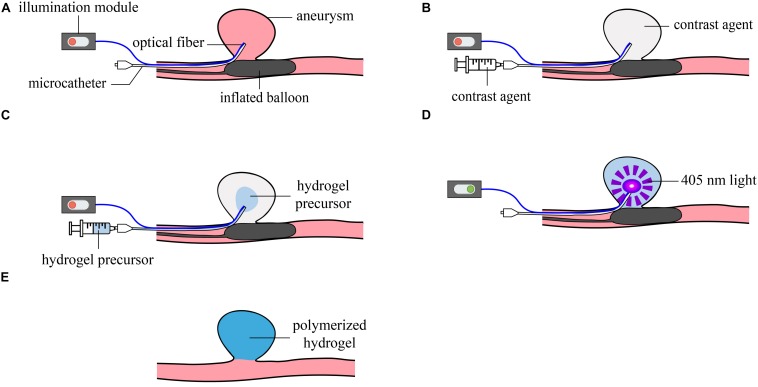
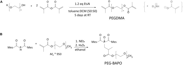
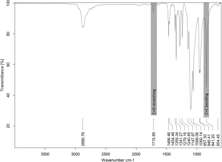
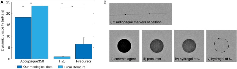
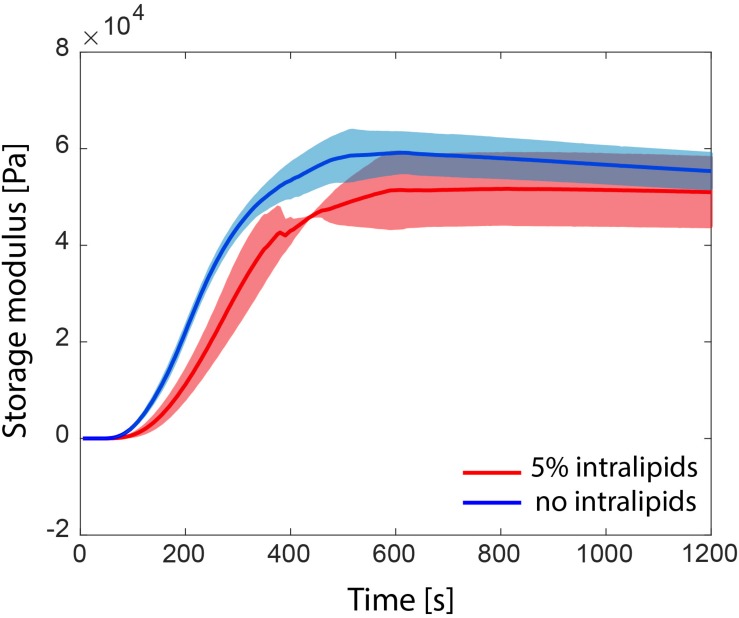
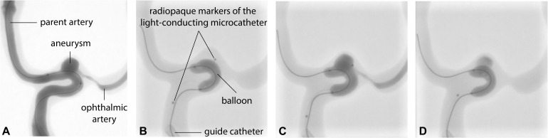
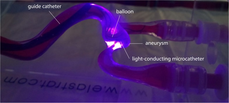
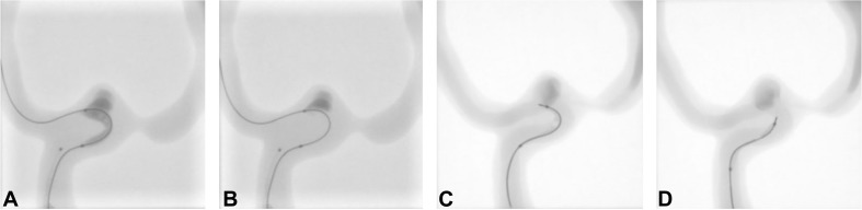
Intracranial aneurysms are increasingly being treated with endovascular therapy, namely coil embolization. Despite being minimally invasive, partial occlusion and recurrence are more frequent compared to open surgical clipping. Therefore, an alternative treatment is needed, ideally combining minimal invasiveness and long-term efficiency. Herein, we propose such an alternative treatment based on an injectable, radiopaque and photopolymerizable polyethylene glycol dimethacrylate hydrogel. The rheological measurements demonstrated a viscosity of 4.86 ± 1.70 mPa.s, which was significantly lower than contrast agent currently used in endovascular treatment (p = 0.42), allowing the hydrogel to be injected through 430 μm inner diameter microcatheters. Photorheology revealed fast hydrogel solidification in 8 min due to the use of a new visible photoinitiator. The addition of an iodinated contrast agent in the precursor contributed to the visibility of the precursor injection under fluoroscopy. Using a customized light-conducting microcatheter and illumination module, the hydrogel was implanted in an in vitro silicone aneurysm model. Specifically, in situ fast and controllable injection and photopolymerization of the developed hydrogel is shown to be feasible in this work. Finally, the precursor and the polymerized hydrogel exhibit no toxicity for the endothelial cells. Photopolymerizable hydrogels are expected to be promising candidates for future intracranial aneurysm treatments.

Keywords: in situ photopolymerization; injectable hydrogels; intracranial aneurysms (IA); light-conducting microcatheter; polyethylene glycol dimethacrylate.


Figures

FIGURE 1
FIGURE 1
Schematic illustration of the different steps of the new treatment concept. (A) Positioning of the light-conducting microcatheter in the aneurysm and inflation of the balloon at the neck; (B) Rinsing of the aneurysm with contrast agent; (C) Injection of the hydrogel precursor; (D) 405 nm illumination and photopolymerization of the hydrogel; (E) Removal of the microcatheter and balloon.
FIGURE 2
FIGURE 2
(A) Synthesis of PEGDMA polymer. (B) Synthesis of PEG-BAPO photoinitiator.
FIGURE 3
FIGURE 3
Schematic illustration of the in vitro setup to evaluate the feasibility of the implantation. The peristaltic pump induced a pulsatile flow of 220 ml/min at 1 Hz. The height of the liquid column ensured a constant pressure of 12 kPa to the system. A parallel bifurcation channel, representing collateral shunting or outflow in the head and neck during balloon inflation at the aneurysm neck was added to dissipate over-pressurization and avoid bursting or displacing the balloon when inflated. The input represents access to the internal carotid artery through which a 9 French introducer sheath and an 8 French guiding catheter were placed, followed by navigation of the light-conducting microcatheter and balloon to the aneurysm site.
FIGURE 4
FIGURE 4
(A) Illumination module to which the optical fibers are connected. (B) Schematic of the light-conducting microcatheter from different views (i) overall, (ii) side, and (iii) front view.
FIGURE 5
FIGURE 5
FT-IR spectrum of PEGDMA polymer.
FIGURE 6
FIGURE 6
(A) Dynamic viscosities for Accupaque350, water, and hydrogel precursor by rheology measurements (n = 3). (B) Radiography snapshot of (i) a deflated balloon with its two proximal and distal radiopaque markers, (ii) pure Accupaque350, (iii) hydrogel precursor, (iv) polymerized hydrogel immediately after photopolymerization and (v) polymerized hydrogel after 36 h-immersion in PBS.
FIGURE 7
FIGURE 7
Storage modulus G’ evolution during the photopolymerization of PEGDMA hydrogels with and without intralipids (n = 3).
FIGURE 8
FIGURE 8
Subtracted (A) and (B–D) unsubtracted fluoroscopic images showing (A) contrast agent injection into the model to visualize the dimensions of the parent artery, aneurysm and ophthalmic artery; (B) guide catheter, inflated balloon (slightly prolapsing in the aneurysm) and light-conducting microcatheter in place; (C) injection of the hydrogel precursor through the microcatheter and (D) polymerized hydrogel after 8 min of 405 nm illumination and removal of the light-conducting microcatheter.
FIGURE 9
FIGURE 9
Optical fiber in situ illumination through the microcatheter in the aneurysm.
FIGURE 10
FIGURE 10
Unsubtracted fluoroscopic images of (A) deflation and (B–D) progressive removal of the balloon. Note that radiopacity is reduced compared to (A) due to slow residual inflow of blood substitute during polymerization. Recycled contrast agent after re-establishment of blood flow throughout the model is visible on (C,D).
FIGURE 11
FIGURE 11
(A) HUVEC viability of different concentrations and dilutions of hydrogel precursors (n = 4). (B) HUVEC viability after 1 week of direct contact with PEGDMA hydrogels either immediately after polymerization or after 1 day of swelling in PBS following polymerization (n = 3).
FIGURE 12
FIGURE 12
Platelet aggregation of different concentrations of hydrogel precursors (n = 3).

References

    1. Abdihalim M., Watanabe M., Chaudhry S. A., Jagadeesan B., Suri M. F. K., Qureshi A. I. (2014). Are coil compaction and aneurysmal growth two distinct etiologies leading to recurrence following endovascular treatment of intracranial aneurysm? J. Neuroimaging 24 171–175. 10.1111/j.1552-6569.2012.00786.x - DOI - PubMed
    1. Avery R. K., Albadawi H., Akbari M., Zhang Y. S., Duggan M. J., Sahani D. V., et al. (2016). An injectable shear-thinning biomaterial for endovascular embolization. Sci. Transl. Med. 8:365ra156. 10.1126/scitranslmed.aah5533 - DOI - PubMed
    1. Barnett P., Hughes A. H., Lin S., Arepally A., Gailloud P. H. (2009). In vitro assessment of Embogel and ultragel radiopaque hydrogels for the endovascular treatment of aneurysms. J. Vasc. Interv. Radiol. 20 507–512. 10.1016/j.jvir.2009.01.005 - DOI - PubMed
    1. Bearat H. H., Preul M. C., Vernon B. L. (2013). Cytotoxicity, in vitro models and preliminary in vivo study of dual physical and chemical gels for endovascular embolization of cerebral aneurysms. J. Biomed. Mater. Res. Part A 101A 2515–2525. 10.1002/jbm.a.34554 - DOI - PubMed
    1. Bonneville F., Sourour N., Biondi A. (2006). Intracranial aneurysms: an overview. Neuroimaging Clin. N. Am. 16 371–382. - PubMed

LinkOut - more resources