Reinforcement of the larynx and trachea in echolocating and non-echolocating bats
- PMID: 32319086
- PMCID: PMC7476186
- DOI: 10.1111/joa.13204
Reinforcement of the larynx and trachea in echolocating and non-echolocating bats
Abstract
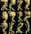
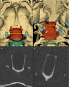
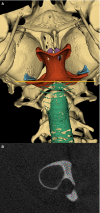
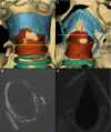
The synchronization of flight mechanics with respiration and echolocation call emission by bats, while economizing these behaviors, presumably puts compressive loads on the cartilaginous rings that hold open the respiratory tract. Previous work has shown that during postnatal development of Artibeus jamaicensis (Phyllostomidae), the onset of adult echolocation call emission rate coincides with calcification of the larynx, and the development of flight coincides with tracheal ring calcification. In the present study, I assessed the level of reinforcement of the respiratory system in 13 bat species representing six families that use stereotypical modes of echolocation (i.e. duty cycle % and intensity). Using computed tomography, the degree of mineralization or ossification of the tracheal rings, cricoid, thyroid and arytenoid cartilages were determined for non-echolocators, tongue clicking, low-duty cycle low-intensity, low-duty cycle high-intensity, and high-duty cycle high-intensity echolocating bats. While all bats had evidence of cervical tracheal ring mineralization, about half the species had evidence of thoracic tracheal ring calcification. Larger bats (Phyllostomus hastatus and Pterpodidae sp.) exhibited more extensive tracheal ring mineralization, suggesting an underlying cause independent of laryngeal echolocation. Within most of the laryngeally echolocating species, the degree of mineralization or ossification of the larynx was dependent on the mode of echolocation system used. Low-duty cycle low-intensity bats had extensively mineralized cricoids, and zero to very minor mineralization of the thyroids and arytenoids. Low-duty cycle high-intensity bats had extensively mineralized cricoids, and patches of thyroid and arytenoid mineralization. The high-duty cycle high-intensity rhinolophids and hipposiderid had extensively ossified cricoids, large patches of ossification on the thyroids, and heavily ossified arytenoids. The high-duty cycle high-intensity echolocator, Pteronotus parnellii, had mineralization patterns and laryngeal morphology very similar to the other low-duty cycle high-intensity mormoopid species, perhaps suggesting relatively recent evolution of high-duty cycle echolocation in P. parnellii compared with the Old World high-duty cycle echolocators (Rhinolophidae and Hipposideridae). All laryngeal echolocators exhibited mineralized or ossified lateral expansions of the cricoid for articulation with the inferior horn of the thyroid, these were most prominent in the high-duty cycle high-intensity rhinolophids and hipposiderid, and least prominent in the low-duty cycle low-intensity echolocators. The non-laryngeal echolocators had extensively ossified cricoid and thyroid cartilages, and no evidence of mineralization/ossification of the arytenoids or lateral expansions of the cricoid. While the non-echolocators had extensive ossification of the larynx, it was inconsistent with that seen in the laryngeal echolocators.
Keywords: Chiroptera; echolocation; mineralization; ossification; respiratory tract.
© 2020 Anatomical Society.
Figures


References
-
- Carpenter, R.E. (1985) Flight physiology of flying foxes, Pteropus poliocephalus . Journal of Experimental Biology, 114, 619–647.
-
- Carpenter, R.E. (1986) Flight physiology of intermediate‐sized fruit bats (Pteropodidae). Journal of Experimental Biology, 120, 79–103.
-
- Carter, R.T. and Adams, R.A. (2014) Ontogeny of the larynx and flight ability in Jamaican fruit bats (Phyllostomidae) with considerations for the evolution of echolocation. Anatomical Record, 297, 1270–1277. - PubMed
-
- Denny, S.P. (1976) Comparative anatomy of the larynx In: The Scientific Basis of Otolaryngology(edsHincliffe, R.andHarrison, D.F.N.). London: Heinemann, pp. 536–545.
Publication types
MeSH terms
LinkOut - more resources
Full Text Sources
Miscellaneous

