. 2020 Jun;24(11):6448-6458.
doi: 10.1111/jcmm.15293. Epub 2020 Apr 21.

Klotho protein contributes to cardioprotection during ischaemia/reperfusion injury

Affiliations

Klotho protein contributes to cardioprotection during ischaemia/reperfusion injury

Agnieszka Olejnik et al. J Cell Mol Med. 2020 Jun.

Abstract

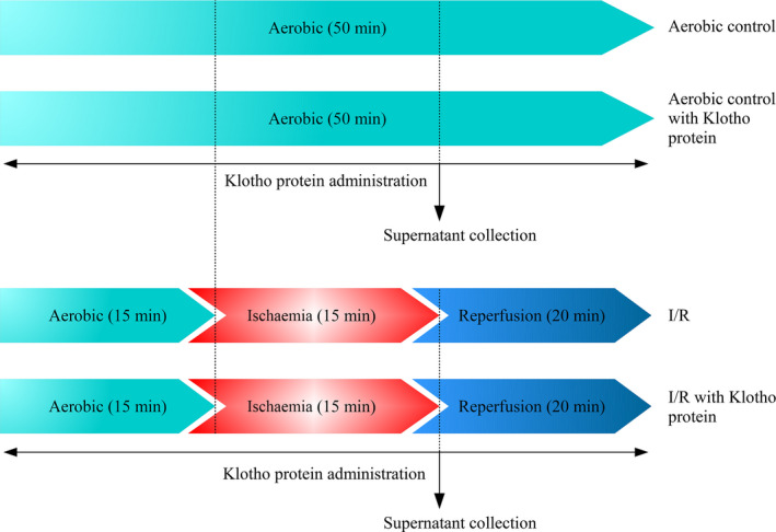
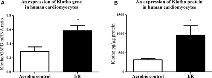
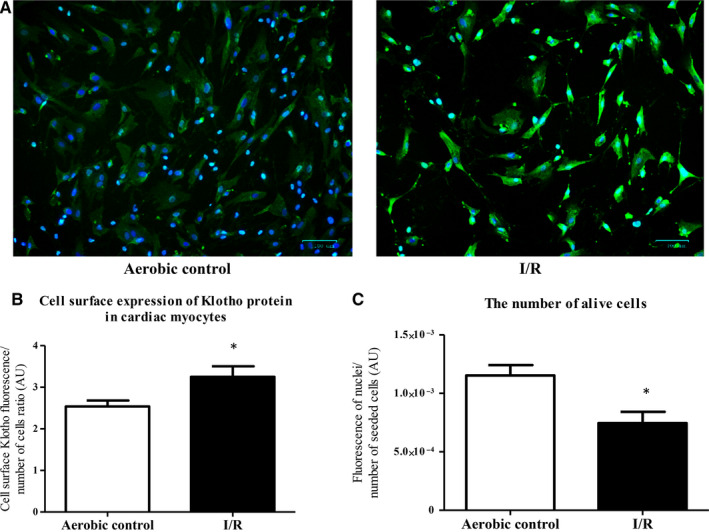
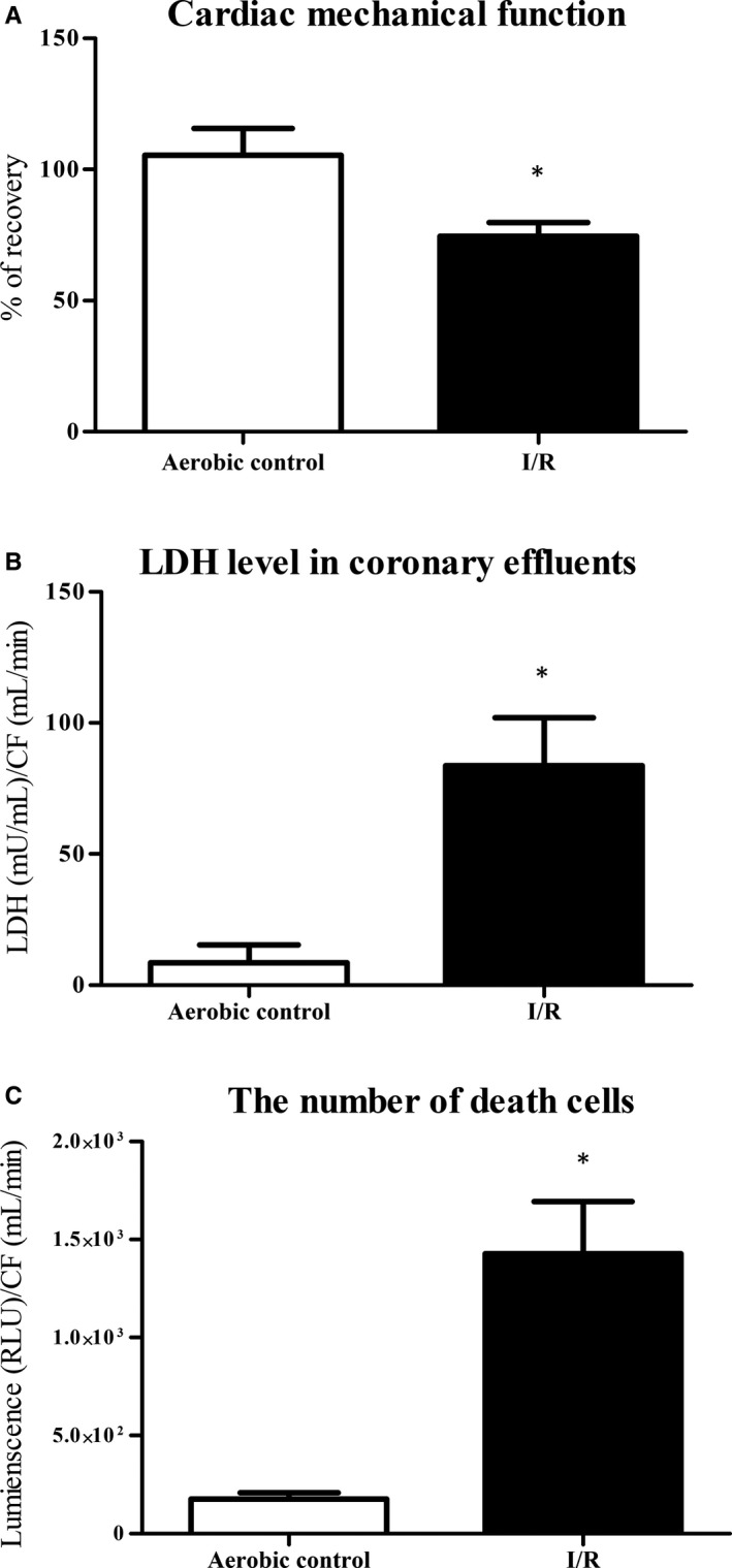
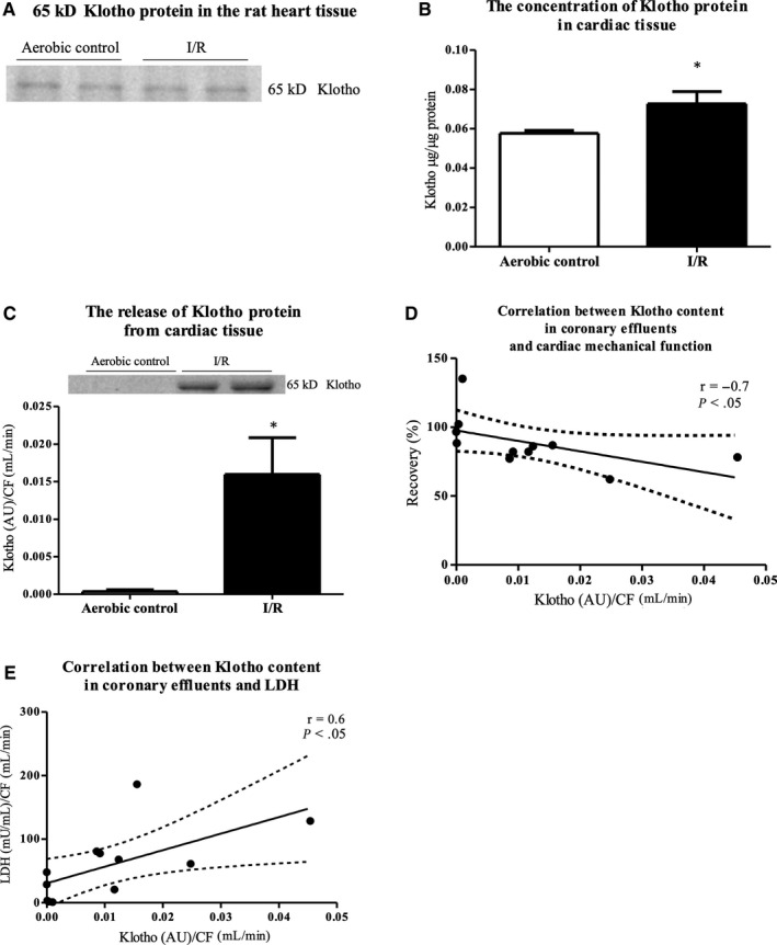
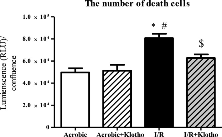
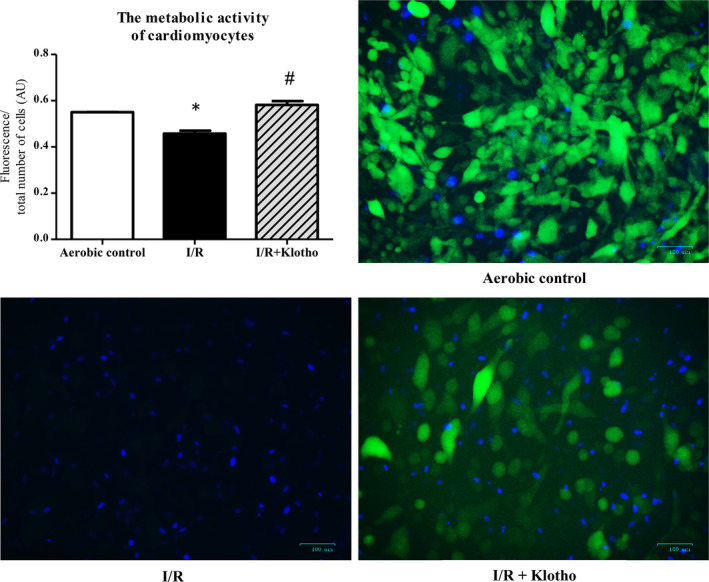
Restoration of blood flow to ischaemic heart inflicts ischaemia/reperfusion (I/R) injury, which manifests in metabolic and morphological disorders. Klotho is a protein with antioxidative and antiapoptotic activity, and is involved in the regulation of inflammation and fibrosis. The aim of the current research was to determine the role of Klotho in the heart subjected to I/R injury, as well as to study Klotho as a potential cardioprotective agent. Human cardiomyocytes and Wistar rat hearts perfused using Langendorff method subjected to I/R have been used. Hemodynamic parameters of heart function, markers of I/R injury, and gene and protein expression of Klotho were measured. Human cardiomyocytes were also incubated in the presence of recombinant Klotho protein, and the viability of cells was measured. There was a higher expression of Klotho gene and protein synthesis in the cardiomyocytes subjected to I/R injury. The compensatory production and release of Klotho protein from cardiac tissue during I/R were also shown. The treatment of cardiomyocytes subjected to I/R with Klotho protein resulted in increased viability and metabolic activity of cells. Thus, Klotho contributes to compensatory mechanism during I/R, and could be used as a marker of injury and as a potential cardiopreventive/cardioprotective agent.

Keywords: Klotho protein; cardiac markers; cardioprotection; ischaemia/reperfusion injury.


Conflict of interest statement

The authors confirm that there are no conflicts of interest.

Figures

FIGURE 1
FIGURE 1
Experimental protocol for in vitro chemical IR injury of cardiomyocytes with and without Klotho administration. I/R, ischaemia/reperfusion
FIGURE 2
FIGURE 2
Klotho in human cardiomyocytes subjected to I/R injury. A, An expression of Klotho gene in human cardiomyocytes was examined by RT‐qPCR and normalized to G6PD, n = 6‐12. B, Quantitative analysis of Klotho protein in human cardiomyocytes by ELISA. Klotho protein concentration was normalized to total protein concentration, n = 18. G6PD, glucose‐6‐phosphate dehydrogenase; I/R, ischaemia/reperfusion; *P < .05 vs aerobic control; all data are expressed as mean ± SEM
FIGURE 3
FIGURE 3
Cell surface expression of Klotho protein in human cardiomyocytes. A, Immunofluorescence staining of human cardiomyocytes for Klotho (green fluorescence) and DAPI for nuclei (blue fluorescence). B, The cell surface expression of Klotho in cardiomyocytes was assessed by measuring the green fluorescence of fixed cells stained with anti‐Klotho antibodies and DyLight 488 and normalized to the total number of cells (blue fluorescence). C, The number of cells was assessed by measuring the fluorescence of cells' nuclei stained by DAPI (blue fluorescence). Graph bars show the average of total fluorescence of cells in each experiment. Magnification 200×; scale bar = 100 μm; AU, arbitrary unit; DAPI, 4',6‐diamidino‐2‐phenylindole; I/R, ischaemia/reperfusion; *P < .05 vs aerobic control; n = 6; all data are expressed as mean ± SEM
FIGURE 4
FIGURE 4
An effect of global I/R injury on isolated rat hearts. A, Recovery of mechanical function of the hearts. Cardiac mechanical function was expressed as the product of heart rate and left ventricular developed pressure—rate pressure product (RPP). RPP was measured at the end of aerobic perfusion (25 min) and at the end of reperfusion (77 min). B, LDH level in coronary effluents as a marker of cell death. LDH level was normalized to CF. C, The number of death cells in rat hearts by luminescent cytotoxicity assay. The data were normalized to CF and expressed in RLU. CF, coronary flow; LDH, lactate dehydrogenase; mU/mL, milli international enzyme units per millilitre; RLU, relative light units; *P < .05 vs aerobic control; mean ± SEM; n = 4‐8
FIGURE 5
FIGURE 5
The expression and release of Klotho protein from the rat heart tissue under I/R injury. A, 65 kDa bands of Klotho protein in the rat heart tissue. Qualitative examination by Western blot. B, The concentration of Klotho protein in the rat heart tissue by ELISA. Klotho protein concentration in tissue homogenates was normalized to total protein concentration. C, Klotho content in coronary effluents (collected at the beginning of reperfusion) by Western blot. The content of Klotho protein in coronary effluents was expressed as AU and normalized to CF. D, Correlation between Klotho content in coronary effluents and cardiac mechanical function and E, LDH level. AU, arbitrary units; CF, coronary flow; LDH, lactate dehydrogenase; mU/mL, milli international enzyme units per millilitre; *P < .05 vs aerobic control; mean ± SEM; n = 4‐8
FIGURE 6
FIGURE 6
An influence of Klotho protein on cell viability. The number of death cells in the presence and absence of recombinant Klotho protein (1 μg/mL) by luminescent cytotoxicity assay. The data were normalized to cell confluence and expressed in RLU. The viability of myocytes was based on the number of death cells. I/R, ischaemia/reperfusion; RLU, relative light units; *P < .05 vs aerobic control; # P < .05 vs aerobic control + Klotho; $ P < .05 vs I/R; mean ± SEM; n = 8
FIGURE 7
FIGURE 7
An influence of Klotho protein on metabolic activity of cardiomyocytes subjected to I/R. Fluorescence microscopy: cells that stained with FDA (green fluorescence) and excluded DAPI (blue fluorescence) were considered viable, whereas cells that excluded FDA and stained with DAPI were considered necrotic. To determine the metabolic activity of cells, green fluorescence (live cells) was divided by the total number of cells (green + blue fluorescence). Graph bars show the average of total fluorescence of cells in each experiment. Magnification 200×; scale bar = 100 μm; AU, arbitrary unit; DAPI, 4',6‐diamidino‐2‐phenylindole; FDA, fluorescein diacetate; I/R, ischaemia/reperfusion; *P < .05 vs aerobic control, # P < .05 vs I/R; mean ± SEM; n = 3‐7

References

    1. Kumar A, Cannon CP. Acute coronary syndromes: diagnosis and management, part I. Mayo Clin Proc. 2010;19:917‐938. - PMC - PubMed
    1. Antman EM, Anbe DT, Armstrong PW, et al. ACC/AHA guidelines for the management of patients with ST‐elevation myocardial infarction—executive summary: a report of the American College of Cardiology/American Heart Association Task Force on Practice Guidelines (Writing Committee to Revise the 1999 Guidelines for the Management of Patients With Acute Myocardial Infarction). Circulation. 2004;110:588‐636. - PubMed
    1. Heusch G, Gersh BJ. The pathophysiology of acute myocardial infarction and strategies of protection beyond reperfusion: a continual challenge. Eur Heart J. 2017;38:774‐784. - PubMed
    1. Zhao Z‐Q, Vinten‐Johansen J. Postconditioning: reduction of reperfusion‐induced injury. Cardiovasc Res. 2006;70:200‐211. - PubMed
    1. Tsutsui H, Kinugawa S, Matsushima S. Oxidative stress and heart failure. Am J Physiol Heart Circ Physiol. 2011;301:H2181‐H2190. - PubMed

Publication types