. 2020 Jul;44(1):14-28.
doi: 10.3892/or.2020.7580. Epub 2020 Apr 9.

HDGF enhances VEGF‑dependent angiogenesis and FGF‑2 is a VEGF‑independent angiogenic factor in non‑small cell lung cancer

Affiliations

HDGF enhances VEGF‑dependent angiogenesis and FGF‑2 is a VEGF‑independent angiogenic factor in non‑small cell lung cancer

Ryoji Eguchi et al. Oncol Rep. 2020 Jul.

Abstract

Non‑small cell lung cancer (NSCLC) accounts for over 80% of all diagnosed lung cancer cases. Lung cancer is the leading cause of cancer‑related deaths worldwide. Most NSCLC cells overexpress vascular endothelial growth factor‑A (VEGF‑A) which plays a pivotal role in tumour angiogenesis. Anti‑angiogenic therapies including VEGF‑A neutralisation have significantly improved the response rates, progression‑free survival and overall survival of patients with NSCLC. However, the median survival of these patients is shorter than 18 months, suggesting that NSCLC cells secrete VEGF‑independent angiogenic factors, which remain unknown. We aimed to explore these factors in human NSCLC cell lines, A549, Lu99 and EBC‑1 using serum‑free culture, to which only EBC‑1 cells could adapt. By mass spectrometry, we identified 1,007 proteins in the culture supernatant derived from EBC‑1 cells. Among the identified proteins, interleukin‑8 (IL‑8), macrophage migration inhibitory factor (MIF), galectin‑1, midkine (MK), IL‑18, galectin‑3, VEGF‑A, hepatoma‑derived growth factor (HDGF), osteopontin (OPN), connective tissue growth factor (CTGF) and granulin (GRN) are known to be involved in angiogenesis. Tube formation, neutralisation and RNA interference assays revealed that VEGF‑A and HDGF function as angiogenic factors in EBC‑1 cells. To confirm whether VEGF‑A and HDGF also regulate angiogenesis in the other NSCLC cell lines, we established a novel culture method. NSCLC cells were embedded in collagen gel and cultured three‑dimensionally. Tube formation, neutralisation and RNA interference assays using the three‑dimensional (3D) culture supernatant showed that VEGF‑A and HDGF were not angiogenic factors in Lu99 cells. By gene microarray in EBC‑1 and Lu99 cells, we identified 61 mRNAs expressed only in Lu99 cells. Among these mRNAs, brain‑derived neurotrophic factor (BDNF), fibroblast growth factor‑2 (FGF‑2) and FGF‑5 are known to be involved in angiogenesis. Tube formation and neutralisation assays clarified that FGF‑2 functions as an angiogenic factor in Lu99 cells. These results indicate that HDGF enhances VEGF‑dependent angiogenesis and that FGF‑2 is a VEGF‑independent angiogenic factor in human NSCLC cells.

Keywords: angiogenesis; fibroblast growth factor-2; hepatoma-derived growth factor; non-small cell lung cancer; vascular endothelial growth factor.


Figures

Figure 1.
Figure 1.
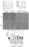
EBC-1 supernatant (sup.) induces angiogenesis. (A) Spheroid-like aggregation and cell shrinkage is induced by serum-free culture in A549 and Lu99 cells, but not in EBC-1 cells. Human NSCLC cell lines, A549, Lu99 and EBC-1, were incubated with or without FBS for 24 h as described in Materials and methods. (B) Flow cytometric analysis using double staining with Annexin V and 7-AAD. EBC-1 cells were incubated in monolayer cultures with or without FBS for 24 h, and then flow cytometric analysis was performed using double staining with Annexin V and 7-AAD as described in Materials and methods. (C) EBC-1 supernatant induces tube formation of HUVECs in 3D culture. HUVECs sandwiched between two layers of collagen were incubated with EBC-1 supernatant at the indicated concentrations for 24 h as described in Materials and methods. ****P<0.001. Each assay was performed in three independent experiments and representative images are shown. Data represent the means ± SEMs of three independent experiments. Statistically significant differences were determined by using one-way factorial analysis of variance (ANOVA)-Dunnett's test. Scale bar, 100 µm. NSCLC, non-small cell lung cancer; HUVECs, human umbilical vein endothelial cells.
Figure 2.
Figure 2.
EBC-1 supernatant (sup.) induces tube formation mediated by both VEGF-A and VEGF-independent angiogenic factors. (A) EBC-1 supernatant transiently induces phosphorylation of VEGFR2 and ERK1/2. HUVECs in collagen-coated culture dishes were incubated with EBC-1 supernatant at 50 µg/ml at the indicated time points in monolayer cultures, and cell extract was then prepared from the cells as described in Materials and methods. (B) VEGF-A neutralisation partially suppresses EBC-1 supernatant-induced tube formation. HUVECs sandwiched between two layers of collagen were incubated with EBC-1 supernatant (50 µg/ml) alone and that together with the mouse monoclonal anti-IgG2B (10 µg/ml) or the anti-VEGF-A antibody for 24 h. ****P<0.001. (C) EBC-1 supernatant-induced phosphorylation of ERK1/2 is partially suppressed by VEGF-A neutralisation. HUVECs in collagen-coated culture dishes were incubated with EBC-1 supernatant (50 µg/ml) alone and that together with the mouse monoclonal anti-IgG2B (10 µg/ml) or the anti-VEGF-A antibody for 5 min. Cell extract was then prepared from the cells. Each assay was performed in three independent experiments and representative images are shown. Data represent the means ± SEMs of three independent experiments. Statistically significant differences were determined by using one-way factorial ANOVA-Tukey's test. Scale bar, 100 µm. VEGF, vascular endothelial growth factor; VEGFR2, vascular endothelial growth factor receptor 2; HUVECs, human umbilical vein endothelial cells.
Figure 3.
Figure 3.
VEGF-A knockdown partially inhibits tube formation induced by EBC-1 supernatant. (A) ELISA for VEGF-A in EBC-1 supernatant. The levels of VEGF-A protein in serum-free culture supernatants (50 µg/ml) of EBC-1 cells and those of the cells transfected with vehicle, siControl or siVEGF-A (#1150) were measured using the Human VEGF-A ELISA Kit as described in Materials and methods. *P<0.05, **P<0.01 and ***P<0.005. (B) VEGF-A in EBC-1 supernatant partially induces tube formation. HUVECs sandwiched between two layers of collagen were incubated with serum-free culture supernatants (50 µg/ml) of EBC-1 cells transfected with vehicle, siControl or siVEGF-A (#1150) for 24 h. ***P<0.005 and ****P<0.001. Each assay was performed in three independent experiments and representative images are shown. Data represent the means ± SEMs of three independent experiments. Statistically significant differences were determined by using one-way factorial ANOVA-Tukey's test. Scale bar, 100 µm. VEGF, vascular endothelial growth factor; HUVECs, human umbilical vein endothelial cells.
Figure 4.
Figure 4.
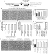
HDGF, but not MK and GRN, is involved in EBC-1 supernatant-induced tube formation. (A) Knockdown of MK, HDGF and GRN by RNAi in the EBC-1 supernatant (sup.). Serum-free culture supernatants (50 µg) of EBC-1 cells transfected with vehicle, siControl, siMK (#706), siHDGF (#724) or siGRN (#619) were used for western blotting. (B) HDGF, but not MK and GRN, is involved in EBC-1 supernatant-induced tube formation. HUVECs sandwiched between two layers of collagen were incubated with serum-free culture supernatants (50 µg/ml) of EBC-1 cells transfected with vehicle, siControl, siMK (#706), siHDGF (#724) or siGRN (#619) for 24 h. ***P<0.005 and ****P<0.001. Each assay was performed in three independent experiments and representative images are shown. Data represent the means ± SEMs of three independent experiments. Statistically significant differences were determined by using one-way factorial ANOVA-Tukey's test. Scale bar, 100 µm. HDGF, hepatoma-derived growth factor; MK, midkine; GRN, granulin; HUVECs, human umbilical vein endothelial cells.
Figure 5.
Figure 5.
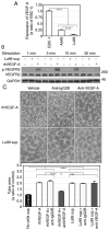
HDGF is directly involved in EBC-1 supernatant-induced tube formation. (A) HDGF knockdown by another HDGF siRNA in the EBC-1 supernatant (sup.). Serum-free culture supernatants (50 µg) of EBC-1 cells transfected with vehicle, siControl or siHDGF (#761) were used for western blotting. (B) HDGF in EBC-1 supernatant partially induces tube formation. HUVECs sandwiched between two layers of collagen were incubated with serum-free culture supernatants (50 µg/ml) of EBC-1 cells transfected with vehicle, siControl or siHDGF (#761) for 24 h. ****P<0.001. (C) rhHDGF partially induces tube formation. HUVECs sandwiched between two layers of collagen were incubated with rhHDGF at 2, 10 and 50 ng/ml or with EBC-1 supernatant (50 µg/ml) for 24 h. *P<0.05, **P<0.01 and ****P<0.001. Each assay was performed in three independent experiments and representative images are shown. Data represent the means ± SEMs of three independent experiments. Statistically significant differences were determined by using One-way factorial ANOVA-Tukey's test. HDGF, hepatoma-derived growth factor; HUVECs, human umbilical vein endothelial cells.
Figure 6.
Figure 6.
VEGF-A and HDGF regulate tube formation induced by the EBC-1 supernatant (sup.). (A and B) Expression of HDGF mRNA in human NSCLC cells. Each total RNA was isolated from EBC-1 cells at the indicated cell densities with or without 10% FBS (A) and from EBC-1, A549 and Lu99 cells incubated with 10% FBS (B) for 24 h. Synthesis of first-strand cDNA and PCR were then performed as described in Materials and methods. The expression levels of HDGF mRNA were normalised to the corresponding levels of GAPDH mRNA (A) and those of 18S-rRNA (B) as an internal control. *P<0.05 and **P<0.01 (B). (C) VEGF-A and HDGF in EBC-1 supernatant induce tube formation. HUVECs sandwiched between two layers of collagen were incubated with serum-free culture supernatants (50 µg/ml) of EBC-1 cells transfected with vehicle, siControl, siHDGF (#724) or siHDGF (#761) together with the mouse monoclonal anti-IgG2B (10 µg/ml) or the anti-VEGF-A antibody. *P<0.05, **P<0.01, ***P<0.005 and ****P<0.001. Each assay was performed in three independent experiments and representative images are shown. Data represent the means ± SEMs of three independent experiments. Statistically significant differences were determined by using one-way factorial ANOVA with Dunnett's test (A and B) and Tukey's test (C) Scale bar, 100 µm. VEGF, vascular endothelial growth factor; NSCLC, non-small cell lung cancer; HDGF, hepatoma-derived growth factor; HUVECs, human umbilical vein endothelial cells.
Figure 7.
Figure 7.
Lu99 supernatant (sup.) induces HDGF-independent tube formation. (A) Procedure of preparation and use of the supernatant derived from Lu99 cells in 3D culture. Lu99 cells embedded in collagen were three-dimensionally incubated with tube-induction medium containing 2% FBS for 24 h, and then Lu99 supernatant was collected as described in Materials and methods. (B) Lu99 supernatant induces tube formation in a cell-density-dependent manner. HUVECs sandwiched between two layers of collagen were incubated with tube-induction medium, in which 3D culture supernatants derived from Lu99 cells cultured at 0–5×10⁶ cells/ml were stratified, for 24 h as described in Materials and methods. *P<0.05, ***P<0.005 and ****P<0.001. (C) Lu99 cell viability in 3D culture was not decreased by HDGF knockdown. Lu99 cells transfected with vehicle, siControl, siHDGF (#724) or siHDGF (#761) were three-dimensionally embedded in collagen and incubated in tube-induction medium for 24 h (a total of 96-h incubations after starting the siRNA transfections) as described in Materials and methods. (D) Protein concentration in Lu99 supernatants. After 24-h incubations in 3D cultures of Lu99 cells transfected with vehicle, siControl, siHDGF (#724) or siHDGF (#761), the culture supernatants derived from these cells were collected and protein concentrations in the supernatants were determined using the Bradford method as described in Materials and methods. (E) ELISA for HDGF in Lu99 supernatant. The levels of HDGF protein in 3D culture supernatants of Lu99 cells transfected with vehicle, siControl, siHDGF (#724) or siHDGF (#761) and that after stratifying vehicle-treated Lu99 supernatant in tube-induction medium were measured using the Human HDGF ELISA Kit as described in Materials and methods. N.D., not detectable. (F) HDGF is not directly involved in Lu99 supernatant-induced tube formation. HUVECs sandwiched between two layers of collagen were incubated with tube-induction medium, in which 3D culture supernatants of Lu99 cells transfected with vehicle, siControl, siHDGF (#724) or siHDGF (#761) were stratified, for 24 h. Each assay was performed in three independent experiments and representative images are shown. Data represent the means ± SEMs of three independent experiments. Statistically significant differences were determined by using one-way factorial ANOVA-Tukey's test. Scale bar, 100 µm. HDGF, hepatoma-derived growth factor; HUVECs, human umbilical vein endothelial cells.
Figure 8.
Figure 8.
Lu99 supernatant (sup.) induces VEGF-independent tube formation. (A) Lu99 cells barely express VEGF-A mRNA. Total RNA was isolated from EBC-1, A549 and Lu99 cells incubated with 10% FBS for 24 h, and then synthesis of first-strand cDNA and PCR was performed. Each level of VEGF-A mRNA was normalised to the corresponding level of 18S-rRNA as an internal control. ****P<0.001. (B) VEGFR2 phosphorylation is not induced by Lu99 supernatant. HUVECs in collagen-coated culture dishes were incubated with tube-induction medium, in which Lu99 supernatant or rhVEGF-A (30 ng/ml) were stratified, at the indicated time points and cell extract was then prepared from the cells. (C) VEGF-A neutralisation failed to suppress Lu99 supernatant-induced tube formation. HUVECs sandwiched between two layers of collagen were incubated with tube-induction medium alone and that together with the mouse monoclonal anti-IgG2B antibody (10 µg/ml) or the anti-VEGF-A antibody, in which either rhVEGF-A (30 ng/ml) or Lu99 supernatant were stratified. ***P<0.005 and ****P<0.001. Each assay was performed in three independent experiments and representative images are shown. Data represent the means ± SEMs of three independent experiments. Statistically significant differences were determined by using one-way factorial ANOVA-Tukey's test. Scale bar, 100 µm. VEGF, vascular endothelial growth factor; HUVECs, human umbilical vein endothelial cells; VEGFR2, vascular endothelial growth factor receptor 2.
Figure 9.
Figure 9.
FGF-2 regulates HDGF- and VEGF-independent tube formation induced by the Lu99 supernatant (sup.). (A) mRNA expressions of BDNF, FGF-2 and FGF-5 in human NSCLC cells. Total RNA was isolated from Lu99, EBC-1 and A549 cells incubated with 10% FBS for 24 h, and then synthesis of first-strand cDNA and PCR was performed. Each expression level of BDNF, FGF-2 and FGF-5 mRNAs was normalised to the corresponding levels of 18S-rRNA as an internal control. ****P<0.001. (B) FGF-2, but not BDNF and FGF-5, is involved in Lu99 supernatant-induced tube formation. HUVECs sandwiched between two layers of collagen were incubated with tube-induction medium alone and that together with the rabbit polyclonal anti-IgG (10 µg/ml), the anti-BDNF, the goat polyclonal anti-IgG (10 µg/ml), the anti-FGF-2 or the anti-FGF-5 antibody, in which Lu99 supernatants were stratified, for 24 h. *P<0.05, ***P<0.005 and ****P<0.001. (C) FGF-2 in Lu99 supernatant induces HDGF- and VEGF-independent tube formation. HUVECs sandwiched between two layers of collagen were incubated with tube-induction medium alone and that together with the mouse monoclonal anti-IgG1 (10 µg/ml) or the anti-FGF-2 antibody, in which Lu99 supernatants were stratified, for 24 h. ****P<0.001. Each assay was performed in three independent experiments and representative images are shown. Data represent the means ± SEMs of three independent experiments. Statistically significant differences were determined by using one-way factorial ANOVA-Tukey's test. Scale bar, 100 µm. FGF-2, fibroblast growth factor-2; HDGF, hepatoma-derived growth factor; VEGF, vascular endothelial growth factor; BDNF, brain-derived neutrophic factor; FGF-2, fibroblast growth factor-2; FGF-5, fibroblast growth factor-5; HUVECs, human umbilical vein endothelial cells.
Figure 10.
Figure 10.
Schematic representation of angiogenic factors in NSCLC cells. HDGF enhances VEGF-dependent angiogenesis in VEGF-expressing NSCLC cells. FGF-2 induces VEGF-independent angiogenesis in VEGF-downregulated NSCLC cells. NSCLC, non-small cell lung cancer; HDGF, hepatoma-derived growth factor; VEGF, vascular endothelial growth factor; FGF-2, fibroblast growth factor-2.

References

    1. Inoue M, Sawada N, Matsuda T, Iwasaki M, Sasazuki S, Shimazu T, Shibuya K, Tsugane S. Attributable causes of cancer in Japan in 2005-systematic assessment to estimate current burden of cancer attributable to known preventable risk factors in Japan. Ann Oncol. 2012;23:1362–1369. doi: 10.1093/annonc/mdr437. - DOI - PubMed
    1. Siegel RL, Miller KD, Jemal A. Cancer statistics, 2016. CA Cancer J Clin. 2016;66:7–30. doi: 10.3322/caac.21332. - DOI - PubMed
    1. Villaruz LC, Socinski MA. The role of anti-angiogenesis in non-small-cell lung cancer: An update. Curr Oncol Rep. 2015;17:26. doi: 10.1007/s11912-015-0448-y. - DOI - PMC - PubMed
    1. Hanahan D, Folkman J. Patterns and emerging mechanisms of the angiogenic switch during tumorigenesis. Cell. 1996;86:353–364. doi: 10.1016/S0092-8674(00)80108-7. - DOI - PubMed
    1. Dvorak HF. Vascular permeability factor/vascular endothelial growth factor: A critical cytokine in tumor angiogenesis and a potential target for diagnosis and therapy. J Clin Oncol. 2002;20:4368–4380. doi: 10.1200/JCO.2002.10.088. - DOI - PubMed

MeSH terms

Substances