

A study analyzing macular microvasculature features after vitrectomy using OCT angiography in patients with idiopathic macular epiretinal membrane
- PMID: 32321458
- PMCID: PMC7178723
- DOI: 10.1186/s12886-020-01429-6
A study analyzing macular microvasculature features after vitrectomy using OCT angiography in patients with idiopathic macular epiretinal membrane
Abstract
Background: To evaluate postoperative changes in retinal capillary plexus and to assess contributing factors in postoperative visual improvement using optical coherence tomography angiography (OCT-A) in patients with idiopathic epiretinal membrane (iERM) post membrane removal.
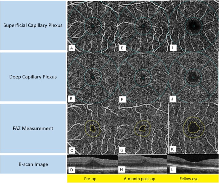
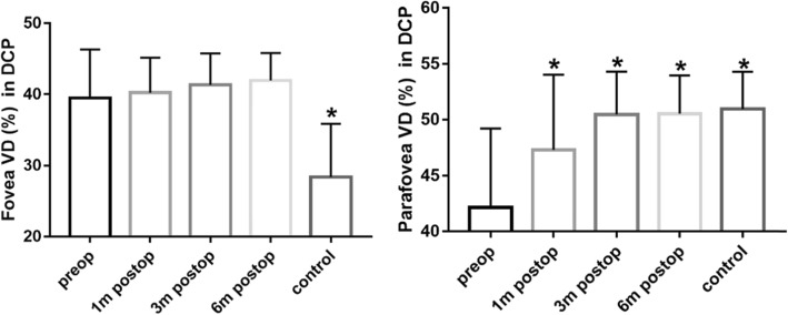
Methods: Patients scheduled for vitrectomy and membrane peel for iERM were enrolled. 35 subjects were included for this study. OCT-A was used to measure the FAZ related parameters and the superficial and deep capillary plexus layers using 3 mm × 3 mm scans. Measurements were taken before surgery and at every post-surgical follow-up. The unaffected fellow eyes were used as controls. Evaluated factors included: BCVA, vessel density (VD) and retinal thickness (RT) in five regions, FAZ area, FAZ perimeter (PERIM), acircularity index (AI) and foveal vessel density (FD).
Results: Compared with the control group, the foveal vessel density (FVD) in superficial capillary plexus (SCP) was greater in the epi-retinal membrane group (P < 0.0001), whereas both groups had comparable parafoveal vessel density (PRVD) in SCP (p > 0.05). After surgery there was a reduction in the PRVD in SCP. The FVD in DCP increased and the PRVD in DCP decreased at baseline (p < 0.001). After surgery there was an increase in PRVD in DCP. By 6 months post-op, the PRVD had no statistically significant difference compared with the control group (p > 0.05). D-value of LogMAR BCVA was positively correlated with pre-op LogMAR BCVA (p < 0.0001), FVD in SCP (p < 0.001). It was negatively correlated with FAZ area (P < 0.001) and PERIM (P < 0.05).
Conclusions: Vitrectomy and membrane removal led to the decrease of VD in SCP and the increase of PRVD in DCP. Patients with a more severe iERM may receive greater visual improvement with surgery.
Trial registration: Trial registration number (TRN) and date of registration. ChiCTR2000031289, retrospectively registered, 2020.03.26.
Keywords: Idiopathic macular epiretinal membrane; Macular microvasculature features; Optical coherence tomography angiography; Retinal thickness; Vitrectomy.
Conflict of interest statement
The authors declare that they have no competing interests.
Figures

References
-
- Inoue M, Arakawa A, Yamane S, Kadonosono K. Long-term outcome of preoperative disrupted inner/outer segment junctions assessed using spectral-domain optical coherence tomography in patients with idiopathic epiretinal membrane. Ophthalmologica. 2012;228(4):222–228. doi: 10.1159/000341606. - DOI - PubMed
MeSH terms
Grants and funding
LinkOut - more resources
Full Text Sources
Miscellaneous
