. 2020 Apr 22;13(1):63.
doi: 10.1186/s13041-020-00601-9.

Arginine is neuroprotective through suppressing HIF-1α/LDHA-mediated inflammatory response after cerebral ischemia/reperfusion injury

Affiliations

Arginine is neuroprotective through suppressing HIF-1α/LDHA-mediated inflammatory response after cerebral ischemia/reperfusion injury

Song-Feng Chen et al. Mol Brain. .

Abstract

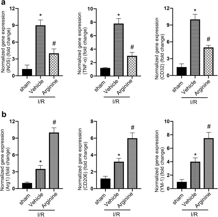
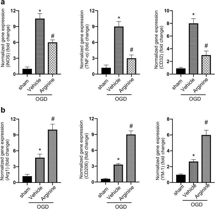
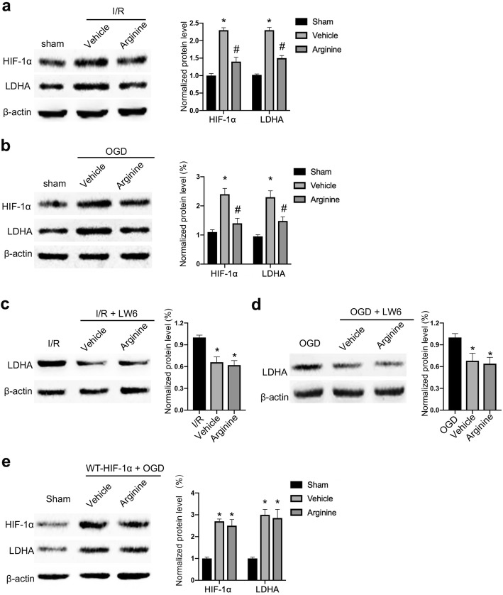
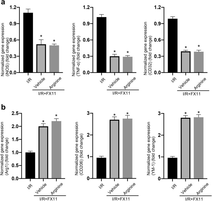
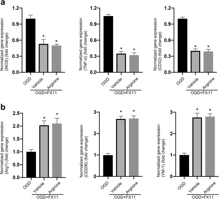
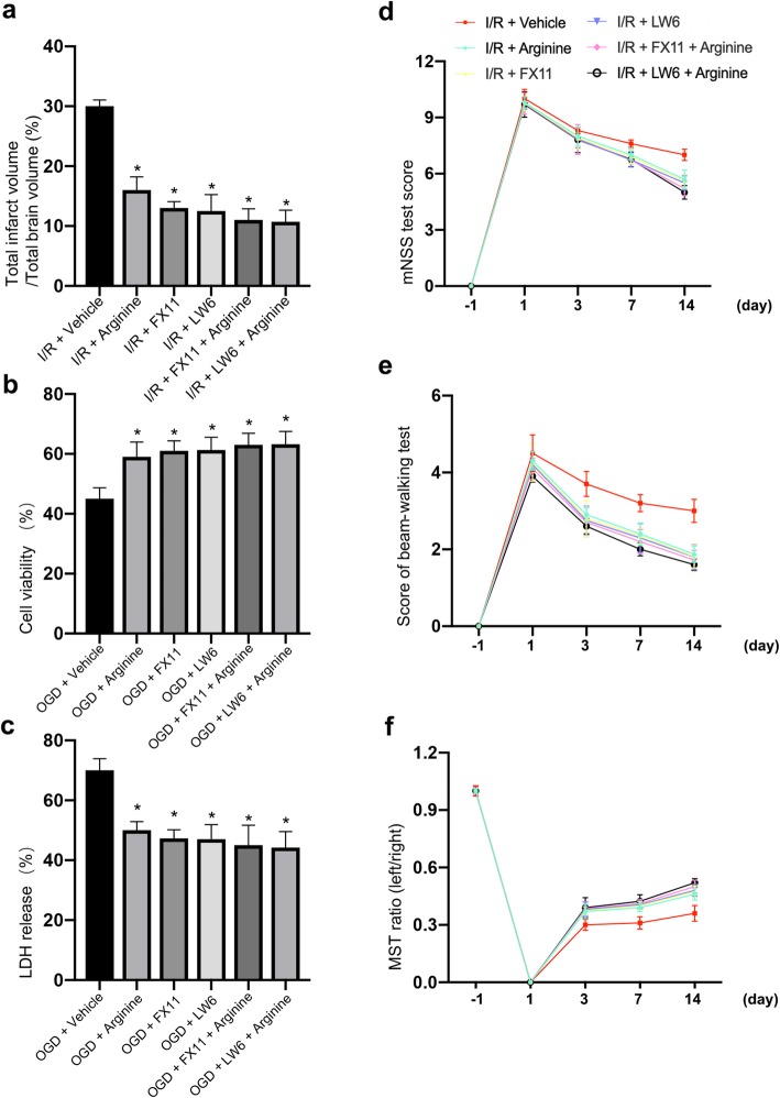
Neuroinflammation is a secondary response following ischemia stroke. Arginine is a non-essential amino acid that has been shown to inhibit acute inflammatory reaction. In this study we show that arginine treatment decreases neuronal death after rat cerebral ischemia/reperfusion (I/R) injury and improves functional recovery of stroke animals. We also show that arginine suppresses inflammatory response in the ischemic brain tissue and in the cultured microglia after OGD insult. We further provide evidence that the levels of HIF-1α and LDHA are increased after rat I/R injury and that arginine treatment prevents the elevation of HIF-1α and LDHA after I/R injury. Arginine inhibits inflammatory response through suppression of HIF-1α and LDHA in the rat ischemic brain tissue and in the cultured microglia following OGD insult, and protects against ischemic neuron death after rat I/R injury by attenuating HIF-1α/LDHA-mediated inflammatory response. Together, these results indicate a possibility that arginine-induced neuroprotective effect may be through the suppression of HIF-1α/LDHA-mediated inflammatory response in microglia after cerebral ischemia injury.

Keywords: Arginine; HIF-1α; Ischemia stroke; LDHA; Neuroinflammation; Neuroprotection.


Conflict of interest statement

The authors declare no competing interests.

Figures

Fig. 1
Fig. 1
Arginine inhibits inflammatory response after ischemia/reperfusion injury. a, b RT-PCR analysis shows pro-inflammation is inhibited and anti-inflammation is enhanced in MCAO rats by arginine. RT-PCR measures the expression level of pro-inflammatory markers, iNOS, TNF-α and CD32 (a) and anti-inflammatory markers, Arg1, YM-1 and CD206 (b) in MCAO rats (n = 6 in each group, *p < 0.05 versus sham, # p < 0.05 versus I/R + vehicle, one-way ANOVA test)
Fig. 2
Fig. 2
Arginine inhibits inflammation response in primary microglia in vitro after OGD insult. a, b RT-PCR analysis shows pro-inflammation is decreased and anti-inflammation is enhanced in OGD microglia by arginine. RT-PCR measures the expression level of pro-inflammatory markers, iNOS, TNF-α and CD32 (a) and anti-inflammatory markers, Arg1, YM-1 and CD206 (b) in OGD microglia (n = 6 in each group, *p < 0.05 versus sham, # p < 0.05 versus I/R + vehicle, one-way ANOVA test)
Fig. 3
Fig. 3
Arginine inhibits HIF-1α to reduce LDHA expression after cerebral I/R injury. a Western blotting shows that HIF-1α and LDHA are raised in MCAO rats and the increase is attenuated by arginine administration (n = 6 in each group, *p < 0.05 versus sham, # p < 0.05 versus vehicle, two-way ANOVA test). b Western blotting shows that HIF-1α and LDHA are raised in in OGD microglia and the increase is attenuated by arginine administration (n = 6 in each group, *p < 0.05 versus sham, # p < 0.05 versus vehicle, two-way ANOVA test). c Western blotting shows that administration of the HIF-1α inhibitor, LW6 decreases the protein level of LDHA in MCAO rats and occludes the effect of arginine (n = 6 in each group, *p < 0.05 versus vehicle, one-way ANOVA test). d Western blotting shows that administration of LW6 decreases the level of LDHA in microglia and occludes the effect of arginine (n = 6 in each group, *p < 0.05 versus OGD, one-way ANOVA test). e Western blotting shows that transfection of WT-HIF-1α abolishes arginine-induced LDHA reduction in OGD BV-2 cells (n = 6 in each group, *p < 0.05 versus OGD, one-way ANOVA test)
Fig. 4
Fig. 4
Arginine inhibits MCAO-induced inflammatory response by the suppression of LDHA. a RT-PCR shows that the increased pro-inflammatory response in MCAO rats is inhibited by FX11. FX11 occludes the pro-inflammation inhibition of arginine (n = 6 in each group, *p < 0.05 versus I/R, one-way ANOVA test). b RT-PCR shows that anti-inflammation is enhanced by FX11 in MCAO rats. FX11 occludes anti-inflammation upregulation by arginine (n = 6 in each group, *p < 0.05 versus I/R, one-way ANOVA test)
Fig. 5
Fig. 5
Arginine inhibits OGD-induced inflammatory response by suppression of LDHA in microglia. a RT-PCR shows that pro-inflammatory response in OGD microglia is inhibited by FX11. FX11 occludes the pro-inflammation inhibition of arginine (n = 6 in each group, *p < 0.05 versus OGD, one-way ANOVA test). b RT-PCR shows that anti-inflammation in OGD microglia is enhanced by FX11. FX11 occludes the anti-inflammation upregulation by arginine (n = 6 in each group, *p < 0.05 versus OGD, one-way ANOVA test)
Fig. 6
Fig. 6
Arginine prevents ischemic neuronal death by inhibiting inflammation response via suppression of HIF-1α/LDHA pathway. a TTC test shows arginine is neuroprotective, and no significant difference in infarct volume among arginine group, FX11 group, LW6 group, FX11 + arginine group and LW6 + arginine group in MCAO rats (n = 6 in each group, *p < 0.05 versus vehicle, one-way ANOVA test). b, c Cell viability test (b) and LDH release (c) show that administration of arginine in OGD microglia attenuates co-cultured neuron death, and no significance among arginine group, FX11 group, LW6 group, FX11 + arginine group and LW6 + arginine group (n = 6 in each group, *p < 0.05 versus vehicle, one-way ANOVA test). d, e, f mNSS test (d), beam-walking test (e), and MST test (f) show that there is no significant difference of functional recovery among arginine group, FX11 group, LW6 group, FX11 + arginine group and LW6 + arginine group (n = 6 in each group, *p < 0.05 versus Vehicle, two-way ANOVA test)

References

    1. Jayaraj RL, Azimullah S, Beiram R, et al. Neuroinflammation: friend and foe for ischemic stroke. J Neuroinflamm. 2019;16(1):142–166. doi: 10.1186/s12974-019-1516-2. - DOI - PMC - PubMed
    1. Lu Y, Li C, Chen Q, et al. Microthrombus-targeting micelles for neurovascular remodeling and enhanced microcirculatory perfusion in acute ischemic stroke. Adv Mater. 2019;31(21):e1808361–e1808373. doi: 10.1002/adma.201808361. - DOI - PubMed
    1. Justicia C, Panés J, Solé S, et al. Neutrophil infiltration increases matrix metalloproteinase-9 in the ischemic brain after occlusion/reperfusion of the middle cerebral artery in rats. J Cereb Blood Flow Metab. 2003;23(12):1430–1440. doi: 10.1097/01.Wcb.0000090680.07515.C8. - DOI - PubMed
    1. Caso JR, Pradillo JM, Hurtado O, et al. Toll-like receptor 4 is involved in brain damage and inflammation after experimental stroke. Circulation. 2007;115(12):1599–1608. doi: 10.1161/CIRCULATIONAHA.106.603431. - DOI - PubMed
    1. Chamorro Á, Dirnagl U, Urra X, et al. Neuroprotection in acute stroke: targeting excitotoxicity, oxidative and nitrosative stress, and inflammation. Lancet Neurol. 2016;15(8):869–881. doi: 10.1016/S1474-4422(16)00114-9. - DOI - PubMed

Publication types

MeSH terms

LinkOut - more resources