. 2020 Apr 22;8(1):58.
doi: 10.1186/s40168-020-00831-y.

Isolation and characterization of diverse microbial representatives from the human skin microbiome

Affiliations

Isolation and characterization of diverse microbial representatives from the human skin microbiome

Collin M Timm et al. Microbiome. .

Abstract

Background: The skin micro-environment varies across the body, but all sites are host to microorganisms that can impact skin health. Some of these organisms are true commensals which colonize a unique niche on the skin, while open exposure of the skin to the environment also results in the transient presence of diverse microbes with unknown influences on skin health. Culture-based studies of skin microbiota suggest that skin microbes can affect skin properties, immune responses, pathogen growth, and wound healing.

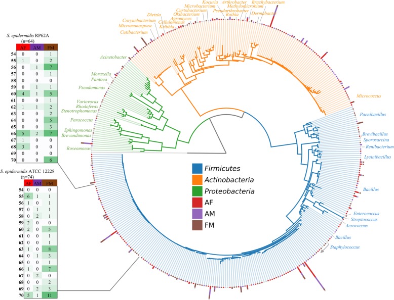
Results: In this work, we greatly expanded the diversity of available commensal organisms by collecting > 800 organisms from 3 body sites of 17 individuals. Our collection includes > 30 bacterial genera and 14 fungal genera, with Staphylococcus and Micrococcus as the most prevalent isolates. We characterized a subset of skin isolates for the utilization of carbon compounds found on the skin surface. We observed that members of the skin microbiota have the capacity to metabolize amino acids, steroids, lipids, and sugars, as well as compounds originating from personal care products.

Conclusions: This collection is a resource that will support skin microbiome research with the potential for discovery of novel small molecules, development of novel therapeutics, and insight into the metabolic activities of the skin microbiota. We believe this unique resource will inform skin microbiome management to benefit skin health. Video abstract.

Keywords: Carbon source utilization; Isolate collection; Skin microbiome.


Conflict of interest statement

The authors declare no competing interests.

Figures

Fig. 1
Fig. 1
Summary of isolation sites and conditions. a Research participants metadata includes age, gender, trans-epidermal water loss, and sebum content. b Example blood agar plate showing colony morphologies. c Total number of isolates by site. d Isolate counts from growth conditions, not including the standard condition of room temperature (RT), blood agar. e Total number of isolates by research participant. Black bars signify research participants for which multiple isolation conditions were performed. See Table S1 for additional metadata
Fig. 2
Fig. 2
Phylogenetic tree of bacterial isolates. 16S tree for bacterial isolates. Sequences with 99% similarity are collapsed into a single node. Concentric columns indicate number of 99% similar 16S sequences by color-coded site. Pop-out tables indicate number of isolates by research participants/site that map to common S. epidermidis strains
Fig. 3
Fig. 3
Bacterial diversity between subjects/sites. Correlation between number of genera by site (raw data shown in a for b antecubital fossa (AF) vs. forearm (AM), c antecubital fossa (AF) vs forehead (FM), and d forearm (AM) vs. forehead (FM). p values were calculated from Pearson correlation with 17 pairs. Outlined bars and points indicate subjects with multiple isolation conditions
Fig. 4
Fig. 4
Fungal isolates. a ITS tree. Blue branches are Ascomycota and orange branches are Basidiomycota. b Isolate FM062_001 identified as Cladosporium caldospoiroides. c Isolate AF056_018 identified as Penicillium dipodomyicola. d Isolate AF064_005 identified as Epicoccum nigrum. e Isolate AF069_007 identified as Naganishia liquefaciens. f Isolate AM063_501 identified as Naganishia liquefaciens. g Isolate AF054_013 identified as Rhodotorula mucilaginosa
Fig. 5
Fig. 5
Skin resource utilization by skin microbiota isolates. a Resource utilization screen. Blue cells indicate a significant color change by the reduction-reporting dye; white cells indicate no significant color change not observed. b Scatterplot of the number of resources found to be utilized by each organism. c Carbon source utilization for generalists across skin isolates is positively related to phylogenetic similarity (four or more carbon sources R2 = 0.169; p = 0.003)

References

    1. Meisel JS, et al. Commensal microbiota modulate gene expression in the skin. Microbiome. 2018;6:20. doi: 10.1186/s40168-018-0404-9. - DOI - PMC - PubMed
    1. Lai Y, et al. Commensal bacteria regulate toll-like receptor 3-dependent inflammation after skin injury. Nat Med. 2009;15:1377–1382. doi: 10.1038/nm.2062. - DOI - PMC - PubMed
    1. Lai Y, et al. Activation of TLR2 by a small molecule produced by Staphylococcus epidermidis increases antimicrobial defense against bacterial skin infections. J. Invest. Dermatol. 2010;130:2211–2221. doi: 10.1038/jid.2010.123. - DOI - PMC - PubMed
    1. Nakatsuji T, et al. Antimicrobials from human skin commensal bacteria protect against <em>Staphylococcus aureus</em> and are deficient in atopic dermatitis. Sci. Transl. Med. 2017;9. - PMC - PubMed
    1. Grice EA, et al. Longitudinal shift in diabetic wound microbiota correlates with prolonged skin defense response. Proc. Natl. Acad. Sci. 2010;107:14799–14804. doi: 10.1073/pnas.1004204107. - DOI - PMC - PubMed

Publication types