. 2020 May 6;40(19):3741-3750.
doi: 10.1523/JNEUROSCI.0046-20.2020. Epub 2020 Apr 22.

Molecular Mechanisms of Non-ionotropic NMDA Receptor Signaling in Dendritic Spine Shrinkage

Affiliations

Molecular Mechanisms of Non-ionotropic NMDA Receptor Signaling in Dendritic Spine Shrinkage

Ivar S Stein et al. J Neurosci. .

Abstract

Structural plasticity of dendritic spines is a key component of the refinement of synaptic connections during learning. Recent studies highlight a novel role for the NMDA receptor (NMDAR), independent of ion flow, in driving spine shrinkage and LTD. Yet little is known about the molecular mechanisms that link conformational changes in the NMDAR to changes in spine size and synaptic strength. Here, using two-photon glutamate uncaging to induce plasticity at individual dendritic spines on hippocampal CA1 neurons from mice and rats of both sexes, we demonstrate that p38 MAPK is generally required downstream of non-ionotropic NMDAR signaling to drive both spine shrinkage and LTD. In a series of pharmacological and molecular genetic experiments, we identify key components of the non-ionotropic NMDAR signaling pathway driving dendritic spine shrinkage, including the interaction between NOS1AP (nitric oxide synthase 1 adaptor protein) and neuronal nitric oxide synthase (nNOS), nNOS enzymatic activity, activation of MK2 (MAPK-activated protein kinase 2) and cofilin, and signaling through CaMKII. Our results represent a large step forward in delineating the molecular mechanisms of non-ionotropic NMDAR signaling that can drive shrinkage and elimination of dendritic spines during synaptic plasticity.SIGNIFICANCE STATEMENT Signaling through the NMDA receptor (NMDAR) is vitally important for the synaptic plasticity that underlies learning. Recent studies highlight a novel role for the NMDAR, independent of ion flow, in driving synaptic weakening and dendritic spine shrinkage during synaptic plasticity. Here, we delineate several key components of the molecular pathway that links conformational signaling through the NMDAR to dendritic spine shrinkage during synaptic plasticity.

Keywords: NMDA receptor; dendritic spine; long-term depression; nitric oxide; structural plasticity; two-photon imaging.


Figures

Figure 1.
Figure 1.
p38 MAPK activity is required for spine shrinkage driven by non-ionotropic NMDAR signaling in response to high-frequency glutamate uncaging. A, Images of dendrites from EGFP-transfected CA1 neurons at 14–18 DIV before and after high-frequency glutamate uncaging (HFU, yellow cross) at individual dendritic spines (yellow arrowhead) in the presence of vehicle, 7-CK (100 μm), and 7-CK with the p38 MAPK inhibitor SB203580 (SB; 2 μm). B, HFU stimulation during vehicle conditions led to long-lasting spine growth (gray filled circles). In the presence of 7-CK, HFU induced dendritic spine shrinkage (red filled circles), which was blocked following inhibition of p38 MAPK activity with SB (black filled circles). The volume of unstimulated neighboring spines (open circles) was unaffected. C, Images of dendrites from EGFP-transfected CA1 neurons at 14–18 DIV before and after HFU (yellow cross) at individual dendritic spines (yellow arrowhead) in the presence of 7-CK with the AMPAR inhibitor NBQX (10 μm), 7-CK with NBQX, and the group I mGluR inhibitors MPEP (15 μm) and CPCCOEt (45 μm), 7-CK in ACSF containing 1 mm Mg2+, or 7-CK with CPP (10 μm). D, Spine shrinkage was induced by HFU in the presence of 7-CK alone (red filled bar; 11 spines/11 cells) and 7-CK with NBQX (blue filled bar; 7 spines/7 cells), NBQX/MPEP/CPCCOEt (purple filled bar; 9 spines/9 cells), or 1 mm Mg2+ (gray filled bar; 6 spines/6 cells), but was blocked by SB203580 (black filled bar; 8 spines/8 cells) and by CPP (brown filled bar; 8 spines/8 cells). *p < 0.05, **p < 0.01, ***p < 0.001, paired two-tailed t test compared with baseline and calculated across cells.
Figure 2.
Figure 2.
Spine shrinkage is induced by HFU in the presence of a lower concentration of L-689,560, which inhibits AMPARs to a lesser extent than 7-CK. A, Left, Representative uEPSCs from individual spines before (gray) and after (red) application of the NMDAR glycine/d-serine site antagonists 7-CK and L-689. Right, Application of 100 μm 7-CK (22 spines/10 cells) greatly reduced and 10 μm L-689 (6 spines/3 cells) partially reduced AMPAR uEPSCs (red filled bars). B, Representative images of dendrites from EGFP-transfected CA1 neurons at 14–18 DIV before and after HFU stimulation (yellow crosses) at individual dendritic spines (yellow arrowheads) in the presence of 100 μm 7-CK or 10 μm L-689. C, HFU stimulation in the presence of 7-CK (black bar; 6 spines/6 cells) or L-689 (red bar; 8 spines/8 cells) caused a stable decrease in spine size at 30 min. Volume of unstimulated neighboring spines (open bars) was not changed. *p < 0.05, **p < 0.01, ***p < 0.001, paired two-tailed t test compared with baseline and calculated across spines in A and cells in C.
Figure 3.
Figure 3.
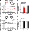
p38 MAPK activity is required for LTD driven by non-ionotropic signaling through the NMDAR. A, Top, Representative uEPSCs from a target spine and an unstimulated neighbor before (light gray) and 25 min after HFU stimulation in the presence of L-689 (target, red; neighbor, dark gray). Bottom, Time course of averaged uEPSC amplitude compared with baseline. B, HFU stimulation in the presence of L-689 induced a long-lasting decrease in uEPSC amplitude of stimulated spines (red line/bar; 7 spines/7 cells), but not of unstimulated neighboring spines (gray line/bar). C, Top, Representative uEPSCs from a target spine and an unstimulated neighbor before (gray) and 25 min after HFU stimulation in the presence of L-689 and the p38 MAPK inhibitor SB (target, black; neighbor, dark gray). Bottom, Time course of averaged uEPSC amplitude compared with baseline. D, The p38 MAPK inhibitor SB blocked LTD induced by HFU in the presence of L-689 (black line/bar; 7 spines/7 cells), while the amplitude of uEPSCs from unstimulated neighboring spines (gray line/bar) did not change. **p < 0.01, ***p < 0.001, paired two-tailed t test compared with baseline and calculated across cells.
Figure 4.
Figure 4.
NOS1AP interaction with nNOS and nNOS enzymatic activity are required for spine shrinkage driven by non-ionotropic signaling through the NMDAR. A, Images of dendrites from EGFP-transfected CA1 neurons at 14–18 DIV before and after high-frequency glutamate uncaging (HFU, yellow cross) at an individual dendritic spine (yellow arrowhead) in the presence of 7-CK (100 μm) and L-TAT-GASA (1 μm) or L-TAT-GESV (1 μm). B, C, Disruption of NOS1AP/nNOS interaction using the active cell-permeant L-TAT-GESV peptide (black filled circles/bar; 15 spines/15 cells), but not the inactive L-TAT-GASA control peptide (red filled circles/bar; 8 spines/8 cells), blocked spine shrinkage induced by non-ionotropic NMDAR signaling. Volume of unstimulated neighboring spines (open circles/bars) was unchanged. D, Images of dendrites from EGFP-transfected CA1 neurons at 14–18 DIV before and after HFU (yellow cross) at an individual dendritic spine (yellow arrowhead) in the presence of 7-CK (100 μm) or 7-CK (100 μm) and L-NNA (100 μm). E, F, Inhibition of NO synthase activity with L-NNA blocked spine shrinkage (solid black circles/bar; 11 spines/11 cells) induced by HFU in the presence of 7-CK (solid red circles/bar; 13 spines/13 cells). The volume of unstimulated neighboring spines (open circles/bars) was unchanged. *p < 0.05, **p < 0.01, ***p < 0.001, paired two-tailed t test compared with baseline and calculated across cells.
Figure 5.
Figure 5.
MK2 activity and cofilin are required for spine shrinkage driven by non-ionotropic NMDAR signaling. A, Images of dendrites from EGFP-expressing neurons (14–18 DIV) showing spine shrinkage (yellow arrowheads) induced by HFU (yellow crosses) in the presence of 7-CK and MK2 inhibitor III (10 μm). B, C, Inhibition of MK2 activity (black filled circles/bar; 11 spines/11 cells) prevented spine shrinkage induced by HFU in the presence of 7-CK (red filled circles/bar; 10 spines/10 cells). Volume of unstimulated neighboring spines (open circles/bars) did not change. D, Images of dendrites from 14–18 DIV neurons expressing CyRFP1 with EGFP and cofilin and ADF shRNAs (KD) or cofilin and ADF shRNAs together with shRNA-resistant cofilin-EGFP (rescue) before and after HFU (yellow crosses) at a single dendritic spine (yellow arrowheads) in the presence of L-689 (10 μm). E, F, KD of cofilin and ADF (black filled circles/bar; 11 spines/11 cells) blocked non-ionotropic NMDAR-dependent spine shrinkage in the presence of L-689 and was rescued by shRNA-resistant cofilin-EGFP (red filled circles/bar; 9 spines/9 cells). Volume of unstimulated neighboring spines (open circles/bars) was not changed. **p < 0.01, ***p < 0.001, paired two-tailed t test compared with baseline and calculated across cells.
Figure 6.
Figure 6.
Non-ionotropic NMDAR-dependent spine shrinkage is associated with loss of cofilin from the spine head. A, Images of CA1 neurons transfected with cofilin shRNAs in combination with shRNA-resistant cofilin-EGFP and CyRFP1 before and after HFU (white crosses) at individual spines (white arrowheads) in the presence of L-689. B, Spine shrinkage (red, CyRFP1) induced by HFU in the presence of L-689 was associated with a decrease of cofilin-GFP protein levels in the spine (green). Cofilin-GFP spine levels were decreased 10 min after HFU in L-689 and stayed decreased until 30 min after HFU in L-689 (16 spines/16 cells). C, Images of dendrites from 14–18 DIV CA1 neurons expressing cofilin shRNAs in combination with shRNA-resistant cofilin-EGFP and CyRFP1 before and after HFU (white crosses) at individual spines (white arrowheads). D, Time course of HFU-induced changes in spine volume (red, CyRFP1) and the amount of cofilin-GFP protein in the spine (green) compared with baseline. Cofilin-GFP spine levels were enriched after HFU and remained enriched for at least 30 min following HFU (6 spines/6 cells). *p < 0.05, **p < 0.01, paired two-tailed t test, comparison of cofilin-EGFP to CyRFP1, calculated across cells.
Figure 7.
Figure 7.
CaMKII activity is required for spine shrinkage driven by non-ionotropic NMDAR signaling. A, Images of dendrites from CA1 neurons of acute slices from P16 to P20 GFP-M mice before and after HFU (yellow cross) at single spines (yellow arrowhead) in the presence of L-689, L-689 with 10 μm KN-62, or L-689 with 5 μm TAT-CN21. B, C, Inhibition of CaMKII activity with KN-62 (red filled circles/bar; 9 spines/9 cells) or TAT-CN21 (gray filled circles/bar; 9 spines/9 cells) blocked spine shrinkage induced by HFU in the presence of L-689 (black filled circles/bar; 8 spines/8 cells). Volume of unstimulated neighboring spines (open bars) was not changed. *p < 0.05, ***p < 0.001, paired two-tailed t test compared with baseline and calculated across cells. D, Proposed model for the non-ionotropic NMDAR signaling pathway that drives spine shrinkage. Glutamate binding to the NMDAR induces conformational changes that drive dendritic spine shrinkage through NOS1AP–nNOS interactions, and the activities of nNOS, p38 MAPK, MK2, CaMKII, and cofilin-dependent severing of the actin cytoskeleton.

Similar articles

Cited by

References

    1. Aow J, Dore K, Malinow R (2015) Conformational signaling required for synaptic plasticity by the NMDA receptor complex. Proc Natl Acad Sci U S A 112:14711–14716. 10.1073/pnas.1520029112 - DOI - PMC - PubMed
    1. Babiec WE, Guglietta R, Jami SA, Morishita W, Malenka RC, O'Dell TJ (2014) Ionotropic NMDA receptor signaling is required for the induction of long-term depression in the mouse hippocampal CA1 region. J Neurosci 34:5285–5290. 10.1523/JNEUROSCI.5419-13.2014 - DOI - PMC - PubMed
    1. Balu DT, Li Y, Puhl MD, Benneyworth MA, Basu AC, Takagi S, Bolshakov VY, Coyle JT (2013) Multiple risk pathways for schizophrenia converge in serine racemase knockout mice, a mouse model of NMDA receptor hypofunction. Proc Natl Acad Sci U S A 110:E2400–E2409. 10.1073/pnas.1304308110 - DOI - PMC - PubMed
    1. Bayer KU, Schulman H (2019) CaM Kinase: still inspiring at 40. Neuron 103:380–394. 10.1016/j.neuron.2019.05.033 - DOI - PMC - PubMed
    1. Birnbaum JH, Bali J, Rajendran L, Nitsch RM, Tackenberg C (2015) Calcium flux-independent NMDA receptor activity is required for Abeta oligomer-induced synaptic loss. Cell Death Dis 6:e1791. 10.1038/cddis.2015.160 - DOI - PMC - PubMed

Publication types

Substances