. 2020 Apr 22;11(1):1941.
doi: 10.1038/s41467-020-15205-z.

The Flemmingsome reveals an ESCRT-to-membrane coupling via ALIX/syntenin/syndecan-4 required for completion of cytokinesis

Affiliations

The Flemmingsome reveals an ESCRT-to-membrane coupling via ALIX/syntenin/syndecan-4 required for completion of cytokinesis

Cyril Addi et al. Nat Commun. .

Abstract

Cytokinesis requires the constriction of ESCRT-III filaments on the side of the midbody, where abscission occurs. After ESCRT recruitment at the midbody, it is not known how the ESCRT-III machinery localizes to the abscission site. To reveal actors involved in abscission, we obtained the proteome of intact, post-abscission midbodies (Flemmingsome) and identified 489 proteins enriched in this organelle. Among these proteins, we further characterized a plasma membrane-to-ESCRT module composed of the transmembrane proteoglycan syndecan-4, ALIX and syntenin, a protein that bridges ESCRT-III/ALIX to syndecans. The three proteins are highly recruited first at the midbody then at the abscission site, and their depletion delays abscission. Mechanistically, direct interactions between ALIX, syntenin and syndecan-4 are essential for proper enrichment of the ESCRT-III machinery at the abscission site, but not at the midbody. We propose that the ESCRT-III machinery must be physically coupled to a membrane protein at the cytokinetic abscission site for efficient scission, uncovering common requirements in cytokinesis, exosome formation and HIV budding.


Conflict of interest statement

The authors declare no competing interests.

Figures
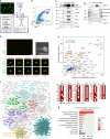
Fig. 1
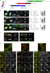
Fig. 1. Proteomics of highly pure and intact post-abscission midbodies revealed known and previously unknown proteins enriched in this organelle.
a Midbody remnant purification. HeLa cells (upper left picture) expressing GFP-MKLP2, a kinesin enriched in midbodies (MB) and midbody remnants (MBRs) were EDTA-treated (Total). After 70g centrifugation, the supernatant (SN) containing MBRs was processed either (1) by differential centrifugations leading to MBR-enriched fraction (MBRE) or (2) subjected to flow cytometry sorting to purify GFP-positive MBRs (MBR+) and their GFP-negative counterpart (MBR−). b Representative pseudo-colored profile of flow cytometry sorting of MBRs. The MBR+ (14% total) and SSC-matched MBR− (44% total) were separated from remaining cells (1%). See Supplementary Fig. 1b. c Western blots of same amounts of protein extracts from Total (Tot), MBR-enriched (MBRE), flow cytometry-sorted MBR− and MBR+ populations. Membranes were blotted repeatedly with indicated antibodies. See also Supplementary Figs. 1c and 6. d Upper left panel: MBR+ population analyzed with cell mask membrane marker. Each individual midbody is positive for GFP-MKLP2 (green) and cell mask (red) Scale bar: 6 μm. Upper right panel: scanning electron microscopy of an isolated MBR. Note the intact and sealed membrane. Lower panels: immunofluorescence stainings of MBR+ for endogenous proteins or membrane marker (red), as indicated. Scale bars: 2 μm. e The Enriched Flemmingsome. Upper panel: merged volcano plot of the mass spectrometry analysis showing the maximum log2(fold change) in x-axis measured between MBR+ and the other fractions (MBRE, MBR−, or Total) and the corresponding –log10(merged p value) in y-axis. color code: proteins significantly enriched in MBR+ when compared with 3 (red), 2 (blue), or 1 (green) of the other fractions. Bottom panel: proteins quantitatively present in MBR+ but not detected in at least two of the other fractions. ALIX (PDCD6IP), syntenin (SDCBP) and syndecan-4 (SDC4) circled in red. f STRING functional association network for the Enriched Flemmingsome. See Supplementary Fig. 3 for details. g GO-term over-representation clusters in the Total Flemmingsome. The size of each bar (x-axis) corresponds to the number of proteins in each cluster and the red gradient the enrichment p values coming from hypergeometric tests. Gray: p value > 0.1.
Fig. 2
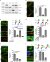
Fig. 2. Syndecan-4, syntenin, ALIX, and CHMP4B colocalize and are highly enriched first at the midbody then at the abscission site.
a The ESCRT-III-ALIX-syntenin-syndecan-4 complex. b Left panels: endogenous localization of ALIX, CHMP4B, syntenin, syndecan-4 (SDC4), and acetylated-tubulin in late bridges displaying abscission site in fixed HeLa cells. The SDC4 antibody recognizes the ectodomain. Middle panels: intensity profiles along the bridge of the corresponding images with matched colors from left panels. Right panels: percentage (mean ± SD) of bridges without and with abscission sites (displaying a pinched tubulin staining on the midbody side) positive for either ALIX, syntenin and syndecan-4. n ≥ 20 cells, N = 3 independent experiments. One-sided Student’s t tests. c Structured illuminated microscopy images of endogenous ALIX/CHMP4B (left) and syntenin/CHMP4B (right), along with acetylated-tubulin staining in late bridges displaying abscission site. Arrows point to ALIX or syntenin localization at the outer rim of the CHMP4B staining. d Snapshots of time-lapse, spinning-disk confocal microscopy movies of cells co-expressing either CHMP4B-GFP/ALIX-mScarlet, GFP-Syntenin/ALIX-mScarlet, or GFP-SDC4/mScarlet-Syntenin. Selected time points show cells (1) before the recruitment of the fluorescently-labeled proteins, (2) after their enrichment at the midbody, (3) after their appearance at the abscission site, and (4) after abscission. Time 0 corresponds to the time frame preceding furrow ingression. See also corresponding Supplementary Movies 2–4. b, d: Scale bars: 10 μm. c Scale bar: 1 μm. Brackets and arrowheads mark the midbody and the abscission site, respectively.
Fig. 3
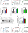
Fig. 3. ALIX directly recruits syntenin and syntenin directly recruits syndecan-4 to the cytokinetic bridge.
a Western blots of protein extracts from cells treated with control, ALIX, syntenin (Synt), or syndecan-4 (SDC4) siRNAs revealed with the indicated antibodies. Loading control: β-tubulin. b Left panels: representative intercellular bridges from control, ALIX or syndecan-4 siRNA-treated cells stained for acetylated-tubulin and endogenous syntenin, as indicated. Right panel: percentage (mean ± SD) of bridges positive for syntenin after control, ALIX and syndecan-4 depletion. n = 31–53 cells, N = 3 independent experiments. (c) Left panels: representative intercellular bridges from control- or ALIX-depleted cells expressing either control (Empty), wild-type ALIX or ALIX F676D mutant (unable to interact with syntenin), and stained for acetylated-tubulin and endogenous syntenin, as indicated. Right panel: percentage (mean ± SD) of bridges with syntenin recruitment in the corresponding conditions. n = 25–31 cells, N = 3 independent experiments. d Left panels: representative intercellular bridges from syntenin-depleted cells expressing either GFP-syntenin wild-type, GFP-syntenin ΔALIX (unable to interact with ALIX) or GFP-syntenin ΔSDC (unable to interact with syndecan-4). Acetylated-tubulin and GFP signals are shown. Right panel: percentage (mean ± SD) of bridges with GFP-tagged syntenin recruitment in the corresponding conditions. n = 14–33 cells, N = 4 independent experiments. e Left panels: representative intercellular bridges from control, ALIX or syntenin siRNA-treated cells and stained for acetylated-tubulin and endogenous syndecan-4, as indicated. Right panel: percentage (mean ± SD) of bridges positive for syndecan-4 after control, ALIX and syntenin depletion. n = 30–40 cells, N = 3 independent experiments. f Left panels: representative intercellular bridges from cells expressing either GFP-syndecan-4 wild-type, GFP-syndecan-4 ΔECD (deleted from its entire extracellular domain) or GFP-syndecan-4 ΔSynt (unable to interact with syntenin). Acetylated-tubulin and GFP signals are shown. Right panel: percentage (mean ± SD) of bridges with GFP-tagged syndecan-4 recruitment in the corresponding conditions. n = 23–41 cells, N = 3 independent experiments. bf Scale bars: 2 μm. Brackets mark the midbody. Panels bf: one-sided Student’s t tests. NS non significant.
Fig. 4
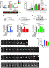
Fig. 4. ALIX, syntenin, and syndecan-4 are required for successful abscission.
a Transduced-HeLa cell lines expressing either GFP, wild-type ALIX-GFP or ALIX F676D-GFP were treated with either control or ALIX siRNAs. Abscission time (from furrow onset to abscission) was determined by phase-contrast time-lapse microscopy. Cumulative plot of the abscission times (upper panels) and mean abscission times (means ± SD) (lower panels) are shown. n = 111–156 cells, N = 3 independent experiments. b Abscission time was determined as above in transduced cell lines expressing either GFP, wild-type mCherry-syntenin, mCherry-syntenin ΔALIX, or mCherry-syntenin ΔSDC and treated with either control or syntenin siRNAs, as indicated. n = 105–147 cells, N = 3 independent experiments. c Abscission time was determined as above in HeLa cells treated with either control or syndecan-4 siRNAs, and transfected with either empty plasmid or plasmid expressing siRNA resistant transcript encoding syndecan-4. n = 130–179 cells, N = 3 independent experiments. Time axis were stopped at 1200 min. d Western blots of protein extracts from cells treated with control, ALIX, syntenin (Synt), syndecan-4 (SDC4), TSG101 siRNAs, or a combination of these siRNAs and revealed with the indicated antibodies. Loading control: calreticulin. eg Abscission time was determined in cells treated with either control, ALIX, syntenin, syndecan-4, TSG101 siRNAs, or a combination of these siRNAs. Mean abscission times (means ± SD) are depicted in (h). n = 120–152 cells, N = 3 independent experiments. All measurements were carried out in parallel but have been displayed in three graphs for clarity. Upper panels ac, eg (time distribution): nonparametric and distribution-free Kolmogorov–Smirnov KS tests. NS nonsignificant. Lower panels ac, h (mean abscission times): one-sided Student’s t tests.
Fig. 5
Fig. 5. Persistent recruitment of ESCRT-III to the abscission site depends on the syndecan4-syntenin-ALIX module.
a Cells treated with the indicated siRNAs were stained for endogenous CHMP4B and acetylated-tubulin. CHMP4B localization in late cytokinetic bridges was classified into three categories: (1) no staining, (2) CHMP4B localized only at the midbody, or (3) CHMP4B localized both at the midbody and at the abscission site (see representative images). The proportion of each category was quantified in control and depleted cells. n = 49–83 cells, N = 3 independent experiments. bd Cells were depleted for either ALIX (b), syntenin (c), or syndecan-4 (d) and transfected with control plasmid (−) or with plasmids encoding either wild type or mutant versions of ALIX, syntenin, or syndecan-4. Upper panels: western blots were revealed with the indicated antibodies. Loading controls: GAPDH or β-tubulin. Lower panels: percentage (mean ± SD) of bridges with CHMP4B at the abscission site in each condition. b n = 28–31 cells; c n = 32–103 cells, d n = 33–56 cells. N = 3 independent experiments. e Cells were treated with control, ALIX, syntenin, syndecan-4, TSG101 siRNAs, alone or in combination. CHMP4B localization was quantified as in (a). n = 26–66 cells, N = 3 independent experiments. fi HeLa cells stably expressing CHMP4B–GFP were treated with either control (f), ALIX (g), syntenin (h), or syndecan-4 (i) siRNAs and recorded by spinning-disk confocal time-lapse microscopy every 10 min. Zooms of the intercellular bridges are displayed. Time 0 corresponds to the frame preceding the arrival of CHMP4B at the midbody. Brackets mark the midbody. Arrows point toward pools of CHMP4B on the midbody side. Red arrows correspond to the CHMP4B leading to abscission (last time frame). Yellow and cyan arrows point to transient and unstable CHMP4B pools observed in depleted cells. See corresponding Supplementary Movies 5–8. j Quantification of cytokinesis with abnormal CHMP4B-GFP behavior (disappearance of signal on the side of the midbody and fragmented cones) for each condition reported in (fi). n = 24–38 cells from 5 independent experiments. One-sided Fisher’s exact tests. a, fi Scale bars: 2 μm. Brackets mark midbodies in (a, fj). Panels ae: means ± SD and one-sided Student’s t tests. NS nonsignificant.
Fig. 6
Fig. 6. Working model: ALIX-syntenin-syndecan-4 couples the ESCRT-III machinery to the plasma membrane at the abscission site for efficient scission.
a ESCRT-III localization at the midbody depends on its recruitment by ALIX (blue) and ESCRT-I/-II (violet), which are targeted to the midbody by MKLP1-associated CEP55 (gray). This first step does not require syntenin or syndecan-4. b ESCRT-III localization at the abscission site, located on one side of the midbody, depends on the tripartite ALIX-syntenin-syndecan-4 module. ALIX-syntenin (red), by directly coupling ESCRT-III (yellow) on the one hand and the transmembrane protein syndecan-4 (green) on the other hand is proposed to help maintain ESCRT-III polymers at the abscission site until the final cut.

Similar articles

Cited by

References

    1. Paweletz N. Walther Flemming: pioneer of mitosis research. Nat. Rev. Mol. Cell Biol. 2001;2:72–75. doi: 10.1038/35048077. - DOI - PubMed
    1. Addi C, Bai J, Echard A. Actin, microtubule, septin and ESCRT filament remodeling during late steps of cytokinesis. Curr. Opin. Cell Biol. 2018;50:27–34. doi: 10.1016/j.ceb.2018.01.007. - DOI - PubMed
    1. Fremont S, Echard A. Membrane traffic in the late steps of cytokinesis. Curr. Biol. 2018;28:R458–R470. doi: 10.1016/j.cub.2018.01.019. - DOI - PubMed
    1. Scourfield EJ, Martin-Serrano J. Growing functions of the ESCRT machinery in cell biology and viral replication. Biochem. Soc. Trans. 2017;45:613–634. doi: 10.1042/BST20160479. - DOI - PubMed
    1. Stoten CL, Carlton JG. ESCRT-dependent control of membrane remodelling during cell division. Semin. Cell Dev. Biol. 2017;74:50–65. doi: 10.1016/j.semcdb.2017.08.035. - DOI - PMC - PubMed

Publication types

MeSH terms