

CRISPR-mediated gene targeting of CK1δ/ε leads to enhanced understanding of their role in endocytosis via phosphoregulation of GAPVD1
- PMID: 32321936
- PMCID: PMC7176688
- DOI: 10.1038/s41598-020-63669-2
CRISPR-mediated gene targeting of CK1δ/ε leads to enhanced understanding of their role in endocytosis via phosphoregulation of GAPVD1
Abstract
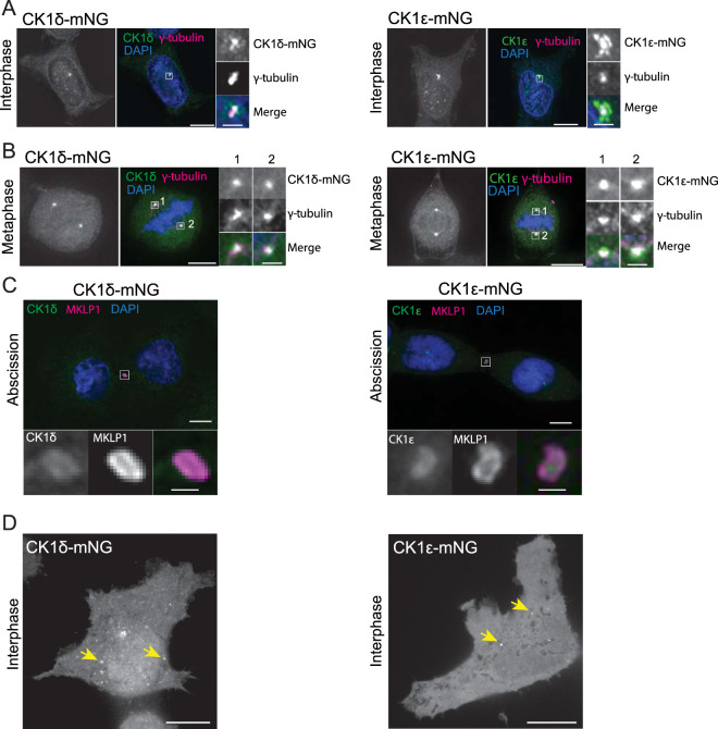
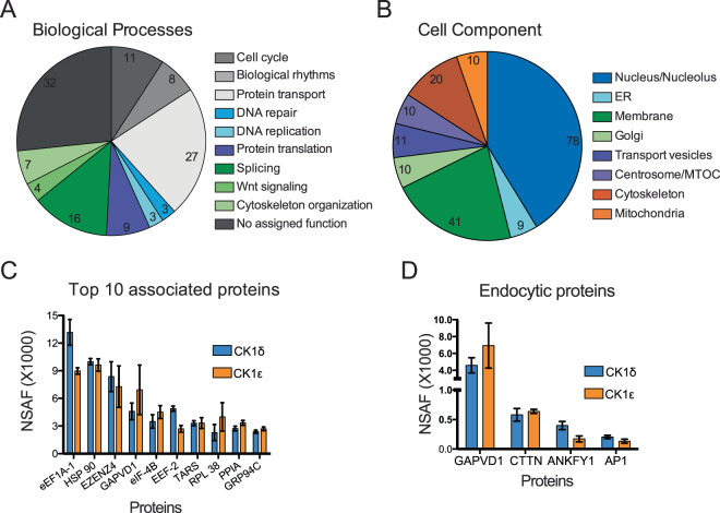
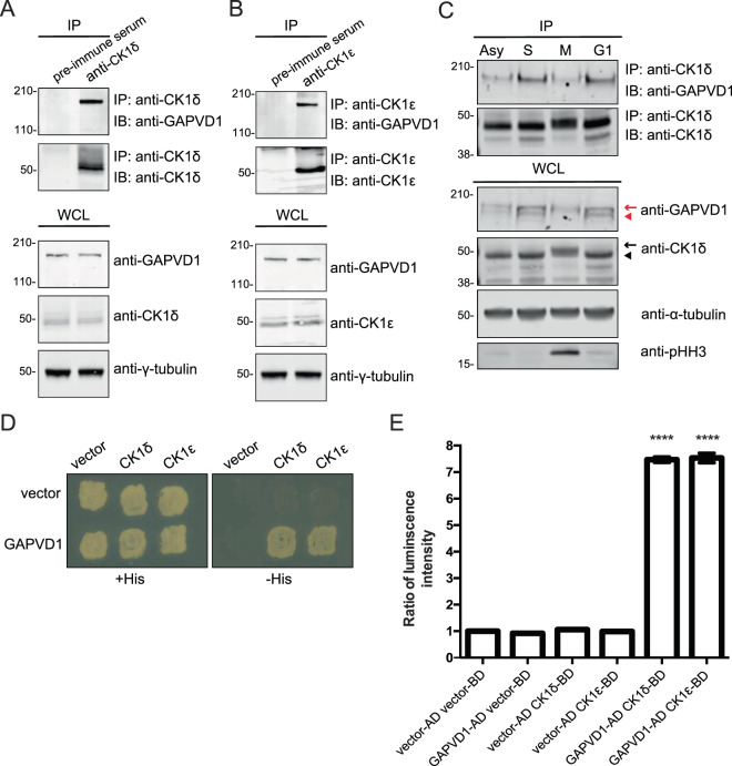
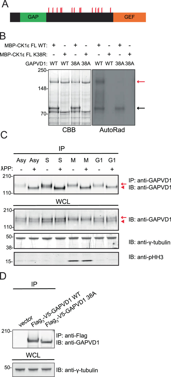
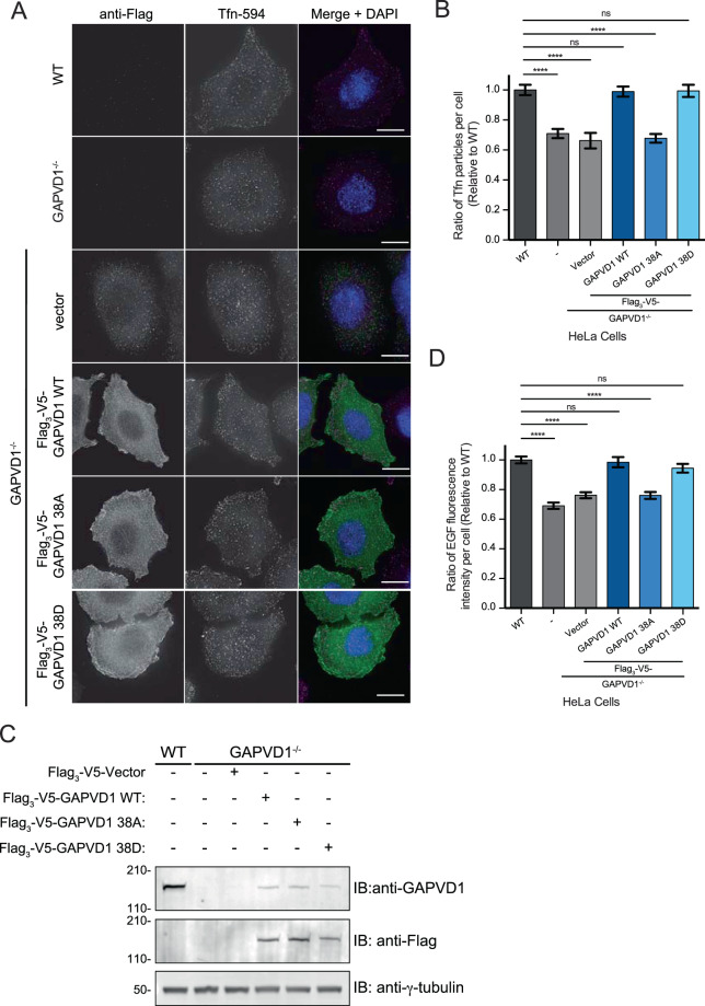
Human casein kinase 1 delta (CK1δ) and epsilon (CK1ε) are members of a conserved family of abundant, ubiquitously expressed serine/threonine kinases that regulate multiple cellular processes including circadian rhythm and endocytosis. Here, we have investigated the localization and interactomes of endogenously tagged CK1δ and CK1ε during interphase and mitosis. CK1δ and CK1ε localize to centrosomes throughout the cell cycle, and in interphase cells to the nucleus, and in both a diffuse and punctate pattern in the cytoplasm. Also, for the first time, they were detected at the midbody during cell division. Mass spectrometry analysis identified a total of 181 proteins co-purifying with a Venus multifunctional (VM)-tagged CK1δ and/or CK1ε. GTPase-activating protein and VPS9 domain-containing protein 1 (GAPVD1), a protein required for efficient endocytosis, was consistently one of the most abundant interacting partners. We demonstrate that GAPVD1 is a substrate of CK1δ/ε with up to 38 phosphorylated residues in vitro and in vivo. Wildtype and a phosphomimetic mutant of GAPVD1, but not a phospho-ablating mutant, were able to rescue defects in transferrin and EGF internalization caused by loss of endogenous GAPVD1. Our results indicate that GAPVD1 is an important interacting partner and substrate of CK1δ/ε for endocytosis.
Conflict of interest statement
The authors declare no competing interests.
Figures
Similar articles
-
Targeting CK1δ and CK1ε as a New Therapeutic Approach for Clear Cell Renal Cell Carcinoma.Pharmacology. 2024;109(6):330-340. doi: 10.1159/000540182. Epub 2024 Jul 17. Pharmacology. 2024. PMID: 38955142
-
Casein kinase 1 delta functions at the centrosome to mediate Wnt-3a-dependent neurite outgrowth.J Cell Biol. 2011 Mar 21;192(6):993-1004. doi: 10.1083/jcb.201011111. J Cell Biol. 2011. PMID: 21422228 Free PMC article.
-
Autokinase Activity of Casein Kinase 1 δ/ε Governs the Period of Mammalian Circadian Rhythms.J Biol Rhythms. 2019 Oct;34(5):482-496. doi: 10.1177/0748730419865406. Epub 2019 Aug 8. J Biol Rhythms. 2019. PMID: 31392916 Free PMC article.
-
Molecular basis for the regulation of the circadian clock kinases CK1δ and CK1ε.Cell Signal. 2017 Feb;31:58-65. doi: 10.1016/j.cellsig.2016.12.010. Epub 2017 Jan 3. Cell Signal. 2017. PMID: 28057520 Review.
-
Casein Kinase 1δ Inhibitors as Promising Therapeutic Agents for Neurodegenerative Disorders.Curr Med Chem. 2022;29(27):4698-4737. doi: 10.2174/0929867329666220301115124. Curr Med Chem. 2022. PMID: 35232339 Review.
Cited by
-
RNA-binding FMRP and Staufen sequentially regulate the Coracle scaffold to control synaptic glutamate receptor and bouton development.Development. 2022 May 1;149(9):dev200045. doi: 10.1242/dev.200045. Epub 2022 May 3. Development. 2022. PMID: 35394012 Free PMC article.
-
Kinase domain autophosphorylation rewires the activity and substrate specificity of CK1 enzymes.Mol Cell. 2022 Jun 2;82(11):2006-2020.e8. doi: 10.1016/j.molcel.2022.03.005. Epub 2022 Mar 29. Mol Cell. 2022. PMID: 35353987 Free PMC article.
-
The DNA Damage Repair Function of Fission Yeast CK1 Involves Targeting Arp8, a Subunit of the INO80 Chromatin Remodeling Complex.Mol Cell Biol. 2024;44(12):562-576. doi: 10.1080/10985549.2024.2408016. Epub 2024 Oct 10. Mol Cell Biol. 2024. PMID: 39387272 Free PMC article.
-
GAPVD1 Promotes the Proliferation of Triple-negative Breast Cancer Cells by Regulating the ERK/MAPK Signaling Pathway.Curr Cancer Drug Targets. 2025;25(5):509-519. doi: 10.2174/0115680096303983240616191051. Curr Cancer Drug Targets. 2025. PMID: 39021189
-
Casein kinase 1 epsilon (CK1ε) as a potential therapeutic target in chronic liver disease.J Vet Sci. 2025 May;26(3):e30. doi: 10.4142/jvs.24321. J Vet Sci. 2025. PMID: 40461423 Free PMC article. Review.
References
Publication types
MeSH terms
Substances
Grants and funding
LinkOut - more resources
Full Text Sources
Research Materials
