Randomized Controlled Trial
. 2020 Apr 22;10(1):6819.
doi: 10.1038/s41598-020-63633-0.

Effect of self-assembling peptide P11-4 on orthodontic treatment-induced carious lesions

Affiliations
Randomized Controlled Trial

Effect of self-assembling peptide P11-4 on orthodontic treatment-induced carious lesions

A Welk et al. Sci Rep. .

Abstract

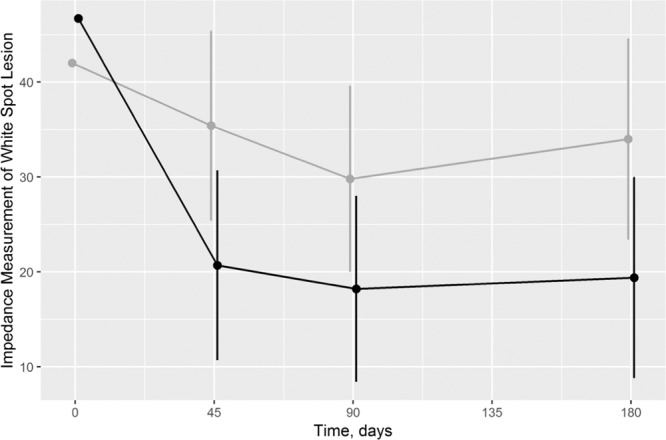
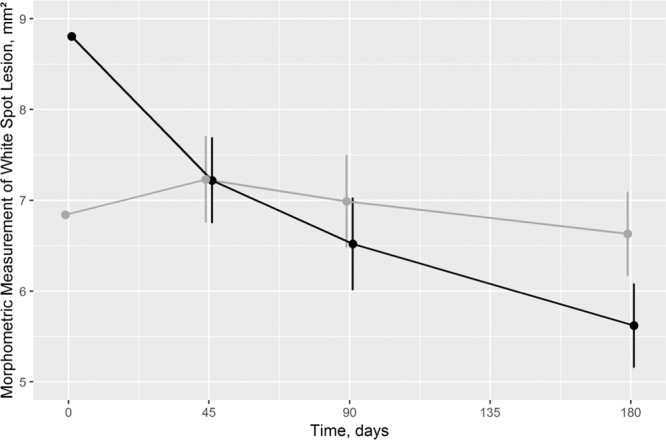
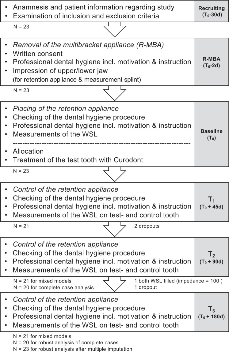
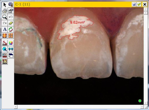
This study aimed to evaluate the effect of self-assembling peptide P11-4 (SAP) in the therapy of initial smooth surface caries (white spot lesions, WSL) following orthodontic multibracket treatment. Twenty-three patients (13f/10m; average age 15.4 years) with at least two teeth with WSL were recruited for the randomised controlled clinical trial with split-mouth design. In opposite to the control teeth, the test teeth were treated with SAP on Day 0. The primary endpoint was the impedance measurement of WSL using customised tray to ensure reproducibility of the measurement location. The secondary endpoint was the morphometric measurement of WSL using a semi-automated approach to determine the WSL size in mm2. Treatment effects were adjusted for site-specific baseline values using mixed models adapted from the cross-over design. Test WSL showed a mean baseline impedance value of 46.7, which decreased to 21.1, 18.4, and 19.7 after 45, 90, and 180 days, respectively. Control WSL showed a mean baseline value of 42.0, which decreased to 35.0, 29.5, and 33.7, respectively. The overall treatment contrast was -13.7 (95% CI: -19.6 - -7.7; p < 0.001). For the secondary endpoint, the test WSL size decreased from 8.8 at baseline to 6.5 after 180 days. The control WSL decreased from 6.8 to 5.7, respectively. The related treatment contrast was -1.0 in favour of test WSL (95% CI: -1.6 - -0.5; p = 0.004). The treatment of initial carious lesions with self-assembling peptide P11-4 leads to superior remineralisation of the subsurface lesions compared with the control teeth.


Conflict of interest statement

The authors declare no competing interests.

Figures

Figure 1
Figure 1
Impedance measurement of White Spot Lesion at different time points (black: test tooth/grey: control tooth). As “temporally and logically, a baseline cannot be a response to treatment, so baseline and response cannot be modeled in an integrated framework”, baseline and response were graphed differently. Consequently, the response and the 95% CI are adjusted for baseline values. p = 0.001, p = 0.005, and p = 0.007 for treatment differences after 45, 90, and 180 days, respectively.
Figure 2
Figure 2
Morphometric measurement of White Spot Lesion Size in mm2 at different time points (black: test tooth/grey: control tooth). As “temporally and logically, a baseline cannot be a response to treatment, so baseline and response cannot be modeled in an integrated framework”, baseline and response were graphed differently. Consequently, the response and the 95% CI are adjusted for baseline values. p = 0.969, p = 0.137, and p = 0.004 for treatment differences after 45, 90, and 180 days, respectively.
Figure 3
Figure 3
Patient Flow Chart.
Figure 4
Figure 4
Image of Impedance measurement of WSL with CarieScan Pro (Orangedental/Biberach/Germany).
Figure 5
Figure 5
Overview image of Morphometric measurement of WSL with Shadepilot (DeguDent/Hanau/Germany).
Figure 6
Figure 6
Computer Screen image of Morphometric measurement of WSL with Shadepilot (DeguDent/Hanau/Germany).

References

    1. Staudt CB, Lussi A, Jacquet J, Kiliaridis S. White spot lesions around brackets: in vitro detection by laser fluorescence. Eur J Oral Sci. 2004;112:237–243. doi: 10.1111/j.1600-0722.2004.00133.x. - DOI - PubMed
    1. Mattousch TJ, van der Veen MH, Zentner A. Caries lesions after orthodontic treatment followed by quantitative light-induced fluorescence: a 2-year follow-up. Eur J Orthod. 2007;29:294–298. doi: 10.1093/ejo/cjm008. - DOI - PubMed
    1. Höchli D, Hersberger-Zurfluh M, Papageorgiou SN, Eliades T. Interventions for orthodontically induced white spot lesions: a systematic review and meta-analysis. Eur J Orthod. 2017;39:122–133. doi: 10.1093/ejo/cjw065. - DOI - PubMed
    1. Ren Y, Jongsma MA, Mei L, van der Mei HC, Busscher HJ. Orthodontic treatment with fixed appliances and biofilm formation–a potential public health threat? Clin Oral Investig. 2014;18:1711–1718. doi: 10.1007/s00784-014-1240-3. - DOI - PubMed
    1. Gorelick L, Geiger AM, Gwinnett AJ. Incidence of white spot formation after bonding and banding. Am J Orthod. 1982;81:93–98. doi: 10.1016/0002-9416(82)90032-X. - DOI - PubMed

Publication types