. 2020 Jul;14(7):1821-1833.
doi: 10.1038/s41396-020-0653-z. Epub 2020 Apr 22.

New virus isolates from Italian hydrothermal environments underscore the biogeographic pattern in archaeal virus communities

Affiliations

New virus isolates from Italian hydrothermal environments underscore the biogeographic pattern in archaeal virus communities

Diana P Baquero et al. ISME J. 2020 Jul.

Abstract

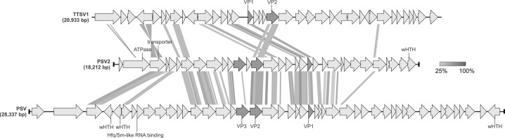
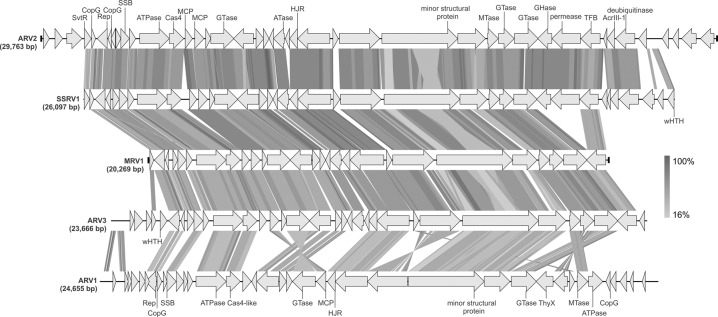
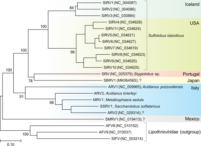
Viruses of hyperthermophilic archaea represent one of the least understood parts of the virosphere, showing little genomic and morphological similarity to viruses of bacteria or eukaryotes. Here, we investigated virus diversity in the active sulfurous fields of the Campi Flegrei volcano in Pozzuoli, Italy. Virus-like particles displaying eight different morphotypes, including lemon-shaped, droplet-shaped and bottle-shaped virions, were observed and five new archaeal viruses proposed to belong to families Rudiviridae, Globuloviridae and Tristromaviridae were isolated and characterized. Two of these viruses infect neutrophilic hyperthermophiles of the genus Pyrobaculum, whereas the remaining three have rod-shaped virions typical of the family Rudiviridae and infect acidophilic hyperthermophiles belonging to three different genera of the order Sulfolobales, namely, Saccharolobus, Acidianus, and Metallosphaera. Notably, Metallosphaera rod-shaped virus 1 is the first rudivirus isolated on Metallosphaera species. Phylogenomic analysis of the newly isolated and previously sequenced rudiviruses revealed a clear biogeographic pattern, with all Italian rudiviruses forming a monophyletic clade, suggesting geographical structuring of virus communities in extreme geothermal environments. Analysis of the CRISPR spacers suggests that isolated rudiviruses have experienced recent host switching across the genus boundary, potentially to escape the targeting by CRISPR-Cas immunity systems. Finally, we propose a revised classification of the Rudiviridae family, with the establishment of six new genera. Collectively, our results further show that high-temperature continental hydrothermal systems harbor a highly diverse virome and shed light on the evolution of archaeal viruses.


Conflict of interest statement

The authors declare that they have no conflict of interest.

Figures

Fig. 1
Fig. 1. Electron micrographs of the VLPs observed in enrichment cultures.
a Fuselloviruses (tailless lemon-shaped virions). b Bicaudaviruses (large, tailed lemon-shaped virions). c Ampullaviruses (bottle-shaped virions). d Rudiviruses (rod-shaped virions). e Lipothrixviruses (filamentous virions). f Globuloviruses (spherical enveloped virions). g Tristromaviruses (filamentous enveloped virions). h Guttaviruses (droplet-shaped virions). Samples were negatively stained with 2% (wt/vol) uranyl acetate. Scale bars: 200 nm.
Fig. 2
Fig. 2. Electron micrographs of the five isolated viruses.
a Metallosphaera rod-shaped virus 1. b Acidianus rod-shaped virus 3. c Saccharolobus solfataricus rod-shaped virus 1. d Pyrobaculum filamentous virus 2. e Pyrobaculum spherical virus 2. Samples were negatively stained with 2% (wt/vol) uranyl acetate. Scale bars: 500 nm; in insets: 100 nm.
Fig. 3
Fig. 3. Genome alignment of the three members of the Globuloviridae family.
The open reading frames (ORFs) are represented by arrows that indicate the direction of transcription. The terminal inverted repeats (TIRs) are denoted by black bars at the ends of the genomes. Genes encoding the major structural proteins are shown in dark gray. The functional annotations of the predicted ORFs are depicted above/below the corresponding ORF. Homologous ORFs and ORF fragments are connected by shading in grayscale based on the level of amino acid sequence identity between the homologous regions. VP, virion protein; wHTH, winged helix-turn-helix domain.
Fig. 4
Fig. 4. Genome comparison of the three members of the Tristromaviridae family.
The open reading frames (ORFs) are represented by arrows that indicate the direction of transcription. The terminal inverted repeats (TIRs) are denoted by black bars at the ends of the genomes. Genes encoding the major structural proteins are shown in dark gray, whereas the four-gene block discussed in the text is shown in black. The functional annotations of the predicted ORFs are depicted above/below the corresponding ORFs. Homologous genes are connected by shading in grayscale based on the level of amino acid sequence identity. The dotted line represents the incompleteness of the TTV1 genome. GHase, glycoside hydrolase; GTase, glycosyltransferase; TP/VP, virion protein.
Fig. 5
Fig. 5. Genome alignment of the Italian rudiviruses.
ARV3, MRV1, and SSRV1 are newly isolated members of the Rudiviridae family, whereas ARV1 and ARV2 were reported previously [49, 68]. The open reading frames (ORFs) are represented by arrows that indicate the direction of transcription. The terminal inverted repeats (TIRs) are denoted by black bars at the ends of the genomes. The functional annotations of the predicted ORFs are depicted above/below the corresponding ORFs. Homologous genes are connected by shading in grayscale based on the amino acid sequence identity. AcrIII-1, anti-CRISPR protein blocking type III CRISPR-Cas systems; ATase, acetyltransferase; CopG, ribbon-helix-helix motif-containing transcription regulator; GHase, glycoside hydrolase; GTase, glycosyltransferase; HJR, Holliday junction resolvase; MCP, major capsid protein; MTase, methyltransferase; Rep, replication initiation protein; SSB, ssDNA binding protein; TFB, Transcription factor B; ThyX, thymidylate synthase; wHTH, winged helix-turn-helix domain.
Fig. 6
Fig. 6. Inferred phylogenomic tree of all known members of the Rudiviridae family based on whole genome VICTOR [59] analysis at the amino acid level.
The tree is rooted with lipothrixviruses, and the branch length is scaled in terms of the Genome BLAST Distance Phylogeny (GBDP) distance formula D6. Only branch support values >70% are shown. For each genome, the abbreviated virus name, GenBank accession number and host organism (when known) are indicated. Question marks denote that the host is not known. The tree is divided into colored blocks according to the geographical origin of the compared viruses.

References

    1. Prangishvili D, Bamford DH, Forterre P, Iranzo J, Koonin EV, Krupovic M. The enigmatic archaeal virosphere. Nat Rev Microbiol. 2017;15:724–39. - PubMed
    1. Munson-McGee JH, Snyder JC, Young MJ. Archaeal viruses from high-temperature environments. Genes (Basel) 2018;9:E128. - PMC - PubMed
    1. Wang H, Peng N, Shah SA, Huang L, She Q. Archaeal extrachromosomal genetic elements. Microbiol Mol Biol Rev. 2015;79:117–52. - PMC - PubMed
    1. Dellas N, Snyder JC, Bolduc B, Young MJ. Archaeal Viruses: Diversity, Replication, and Structure. Annu Rev Virol. 2014;1:399–426. - PubMed
    1. Wang H, Guo Z, Feng H, Chen Y, Chen X, Li Z, et al. Novel Sulfolobus virus with an exceptional capsid architecture. J Virol. 2018;92:e01727–17. - PMC - PubMed

Publication types