. 2020 Apr 22;10(1):6839.
doi: 10.1038/s41598-020-63913-9.

Computer-aided synthesis of dapsone-phytochemical conjugates against dapsone-resistant Mycobacterium leprae

Affiliations

Computer-aided synthesis of dapsone-phytochemical conjugates against dapsone-resistant Mycobacterium leprae

Shasank S Swain et al. Sci Rep. .

Abstract

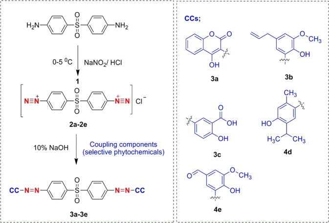
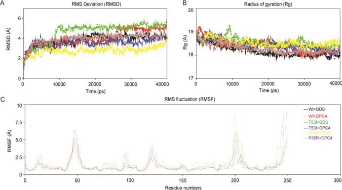
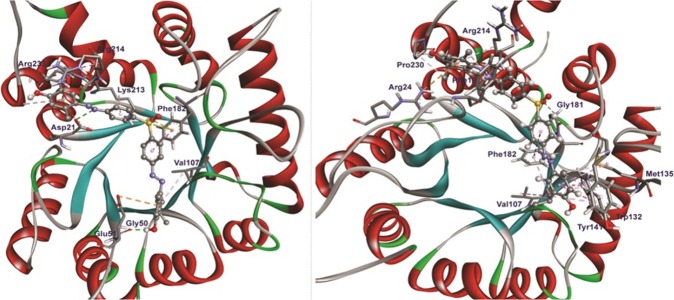
Leprosy continues to be the belligerent public health hazard for the causation of high disability and eventual morbidity cases with stable prevalence rates, even with treatment by the on-going multidrug therapy (MDT). Today, dapsone (DDS) resistance has led to fear of leprosy in more unfortunate people of certain developing countries. Herein, DDS was chemically conjugated with five phytochemicals independently as dapsone-phytochemical conjugates (DPCs) based on azo-coupling reaction. Possible biological activities were verified with computational chemistry and quantum mechanics by molecular dynamics simulation program before chemical synthesis and spectral characterizations viz., proton-HNMR, FTIR, UV and LC-MS. The in vivo antileprosy activity was monitored using the 'mouse-foot-pad propagation method', with WHO recommended concentration 0.01% mg/kg each DPC for 12 weeks, and the host-toxicity testing of the active DPC4 was seen in cultured-human-lymphocytes in vitro. One-log bacilli cells in DDS-resistant infected mice footpads decreased by the DPC4, and no bacilli were found in the DDS-sensitive mice hind pads. Additionally, the in vitro host toxicity study also confirmed that the DCP4 up to 5,000 mg/L level was safety for oral administration, since a minor number of dead cells were found in red color under a fluorescent microscope. Several advanced bioinformatics tools could help locate the potential chemical entity, thereby reducing the time and resources required for in vitro and in vitro tests. DPC4 could be used in place of DDS in MDT, evidenced from in vivo antileprosy activity and in vitro host toxicity study.


Conflict of interest statement

The authors declare no competing interests.

Figures

Figure 1
Figure 1
Diagrammatic presentation of the mutation event of MlDHPS enzyme at folP1 site confirming DDS resistance. At codons 53 and 55, the enzyme mutates sequences coding Ala or Ile or Val and Arg or Leu, respectively.
Figure 2
Figure 2
Stepwise synthesis process of DDS-phytochemical conjugates (DPCs).
Figure 3
Figure 3
Conformational stability of MlDHPS‐DDS/DPC4 complexes during 40‐ns time period indicated in different color graph. (A) RMSD of MlDHPS‐DDS/DPC4 complexes; (B) Rg of MlDHPS‐DDS/DPC4 complexes; (C) Cα‐RMSF profile of the target, MlDHPS‐DDS/DPC4 complexes during 40 nano second molecular dynamic simulation.
Figure 4
Figure 4
(A) Stability of MlDHPS‐DDS/DPC4 complexes inferred through intermolecular Hydrogen‐bonds; (B) Principal component analysis of the DCP4/DDS interacted with the target enzyme, MlDHPS of both wild‐type and mutant‐type structures. Eigenvalues for the MlDHPS‐DCP4/DDS bound models as a function of eigenvector and the plot displays the eigenvalues of only the first 20 eigenvectors; and (C) The cloud represents the 40‐ns trajectories projected onto the first two eigenvectors, EV1 and EV2. The X‐axis and Y‐axis show the projection of the structures of the backbone atoms in the molecular dynamics trajectories of complexes onto the phase space defined.
Figure 5
Figure 5
Molecular interactions of DPC4 with mutant MlDHPS (P55R) during docking. The interactions image was generated using BIOVIA DSV and interacted amino acids are highlighted.
Figure 6
Figure 6
The log10 concentration values were determined taking probit points of cytotoxicity ascertained by AO/EB staining and MTT assay; and probits were plotted for DPC4 during toxicity study with lymphocytes, for establishing toxicity.

References

    1. Swain SS, et al. Molecular docking and simulation study for synthesis of alternative dapsone derivative as a newer anti-leprosy drug in multidrug therapy. J. Cellular. Biochem. 2018;119:9838–52. doi: 10.1002/jcb.27304. - DOI - PubMed
    1. Chaitanya SV, Das M, Bhat P, Ebenezer M. Computational modelling of dapsone interaction with dihydropteroate synthase in Mycobacterium leprae; insights into molecular basis of dapsone resistance in leprosy. J. Cell. Biochem. 2015;116:2293–2303. doi: 10.1002/jcb.25180. - DOI - PubMed
    1. World Health Organization Weekly Epidemiological Record. 2016;92:501–20.
    1. Head MG, et al. Research investments in global health: A systematic analysis of UK infectious disease research funding and global health metrics, 1997-2013. EBioMedicine. 2015;3:180–90. doi: 10.1016/j.ebiom.2015.12.016. - DOI - PMC - PubMed
    1. Saunderson PR. Drug-resistant Mycobacterium leprae. Clin. Dermatol. 2016;34:79–81. doi: 10.1016/j.clindermatol.2015.10.019. - DOI - PubMed

Publication types

MeSH terms