. 2020 Mar 29:24:139-148.
doi: 10.1016/j.jare.2020.03.009. eCollection 2020 Jul.

The landscape of immune cell infiltration and its clinical implications of pancreatic ductal adenocarcinoma

Affiliations

The landscape of immune cell infiltration and its clinical implications of pancreatic ductal adenocarcinoma

Caiming Xu et al. J Adv Res. .

Abstract

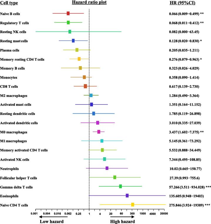
The details of the immunological microenvironment and its clinical implications for pancreatic cancer are still unclear. In this study, we obtained data from public databases, such as the Gene Expression Omnibus, the Cancer Genome Atlas Program, the International Cancer Genome Consortium Data Portal, the ArrayExpress Data Warehouse, and the cBioPortal for Cancer Genomics. We used these data to evaluate the pattern of immune cells infiltration in pancreatic ductal adenocarcinoma (PDAC) tissues. We observed that the levels of M0 macrophages and activated dendritic cells in tumor tissues were significantly higher than that in para-tumor tissues. M0 macrophages, gamma delta T cells and naive CD4 T cells were independent predictive factors of a poor outcome for PDAC patients. An immune score determined by M0 macrophages, gamma delta T cells and naive CD4 T cells could predict the survival of patients. The results of this study suggest that the infiltration of immune cells, such as M0 macrophages, may be a possible target for the treatment of PDAC. However, these findings need to be confirmed by additional studies.

Keywords: Immune cell infiltration; M0 macrophages; Pancreatic ductal adenocarcinoma; Prognosis.


Conflict of interest statement

The authors have declared no conflict of interest

Figures

None
Graphical abstract
Fig. 1
Fig. 1
Flowchart of the study. GEO: Gene Expression Omnibus (https://www.ncbi.nlm.nih.gov/geo/). TCGA: The Cancer Genome Atlas Program (https://portal.gdc.cancer.gov/). ICGC: International Cancer Genome Consortium Data Portal (https://icgc.org/). ArrayExpress: ArrayExpress Data Warehouse (https://www.ebi.ac.uk/arrayexpress/). cBioPortal: cBioPortal for Cancer Genomics (http://www.cbioportal.org/).
Fig. 2
Fig. 2
The levels of immune cells in PDAC tissues and para-PDAC tissues. Two GEO series, GSE62452 (N = 22 pairs) and GSE28735 (N = 23 pairs), were used to evaluate the pattern of immune cell infiltration between PDAC tissue and para-tumor tissue (A). The levels of M0 macrophages (M0) and activated dendritic cells (ADCs) in tumor tissues were significantly high than that in para-tumor tissues. However, the level of naive B cells was significantly reduced (B). The multiple logistic regression demonstrated M0 and ADC were the independent predictors of PDAC and a predictive score was determined by M0 and ADC (C). The ROC curve suggested that M0 in combination with ADC could significantly distinct PDAC from non-PDAC (D). Wilcoxon test and P < 0.05 indicates significantly difference for Fig. 2B.
Fig. 3
Fig. 3
The univariate cox proportional hazards regression model of immune cell infiltration. In the training cohort, we observed that Naive B cells, regulatory T cells, resting mast cells, and memory resting CD4 T cells significantly decreased the hazard ratio for death. However, M0 macrophages, gamma delta T cells and naive CD4 T cells significantly increased the hazard ratio for death. * indicates P < 0.05, ** indicates P < 0.01. ***indicates P < 0.001.
Fig. 4
Fig. 4
Evaluating the proportional hazards assumption of multiple Cox regression. The schoenfeld residual of naive B cells (A), regulatory T cells (B), resting mast cells (C), memory resting CD4 T cells (D), M0 macrophages (E), gamma delta T cells (F), and naive CD4 T cells (G) were not dependent on the time. This suggests that the assumption of multiple Cox regression is satisfied.
Fig. 5
Fig. 5
Development and validation of immune score for diagnosis of PDAC. The multiple cox proportional hazards regression suggested that M0 macrophages, naive CD4 T cells and gamma delta T cells were independent risk factors of survival, and an immune score was developed based on these variables (A). The optimal cut-off of this index was 0.4, which was determined by X-title (B and C). The Kaplan-Meier curve and log-rank test suggested that the survival of patients with an immune score no greater than 0.4 was significantly longer than the survival of those with an immune score greater than 0.4 in training cohort (D), validation cohort (E), and the entire cohort (F). In addition, and the relapse-free survival (RFS) time of patients whose immune score was no greater than 0.4 was longer than that of patients with an immune score greater than 0.4 (G). The prognostic power of immune score was significantly superior to the TNM stage in both the training cohort and the validation cohort (H).
Fig. 6
Fig. 6
Gene set enrichment analysis (GSEA) of PDAC with different immune score. 122 samples from TCGA were divided into two groups, the immune score ≤ 0.4 group (N = 96) and the immune score greater than 0.4 group (N = 26). PDAC patients with immune score >0.4 have a low enrichment score for the following biological processes of cell chemotaxis (A), leukocyte chemotaxis (B) and chemokine mediated signaling pathways (C). The expression levels of chemokine (C-X-C motif) ligand 9 (CXCL9), CXCL10, CXCL11, CXCL13, chemokine (C-C motif) ligand 15 (CCL15), CCL17, chemokine (C-X-C motif) receptor 2 (CXCR2), and CXCR6 were significantly decreased in patients with an immune score >0.4, * indicates P < 0.05 , ** indicates P < 0.01. ***indicates P < 0.001 (D). PDAC patients with immune score >0.4 have a low enrichment score for the following biological processes of activation of immune response (E), immune response regulating cell surface receptor signaling pathway (F), antigen receptor mediated signaling pathway (G), natural killer cell activation (H), dendritic cell migration (I) and the molecular function of cytokine receptor activity (J).

References

    1. Siegel R.L., Miller K.D., Jemal A. Cancer statistics, 2019. CA Cancer J Clin. 2019;69(1):7–34. - PubMed
    1. Zhang X., Kumstel S., Jiang K., Meng S., Gong P., Vollmar B. LW6 enhances chemosensitivity to gemcitabine and inhibits autophagic flux in pancreatic cancer. J Adv Res. 2019;20:9–21. - PMC - PubMed
    1. Kamisawa T., Wood L.D., Itoi T., Takaori K. Pancreatic cancer. Lancet. 2016;388(10039):73–85. - PubMed
    1. Watt J., Kocher H.M. The desmoplastic stroma of pancreatic cancer is a barrier to immune cell infiltration. Oncoimmunology. 2013;2(12):1–3. - PMC - PubMed
    1. Fan J.Q., Wang M.F., Chen H.L., Shang D., Das J.K., Song J. Current advances and outlooks in immunotherapy for pancreatic ductal adenocarcinoma. Mol Cancer. 2020;19(1):1–22. - PMC - PubMed