. 2020 Mar 10:19:100167.
doi: 10.1016/j.pacs.2020.100167. eCollection 2020 Sep.

Side-viewing photoacoustic waveguide endoscopy

Affiliations

Side-viewing photoacoustic waveguide endoscopy

Christopher Miranda et al. Photoacoustics. .

Abstract

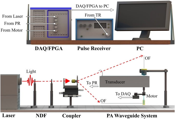
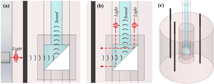
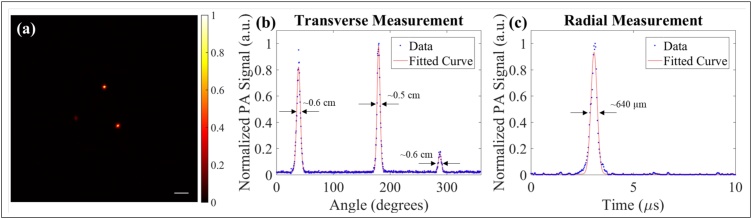
Side-viewing hollow optical waveguides allow for minimally invasive endoscopy by concentrically guiding light and sound for photoacoustic generation and detection. Here, we characterize the side-viewing photoacoustic waveguide (PWG) endoscope by scanning 7.2 μm diameter carbon fiber threads within phantom tissues and animal tissues. Photoacoustic signals are carried along the 5.5 and 10.0 cm length of the PWG with minimal attenuation. Thus, this technology enables 360°, deep-tissue photoacoustic imaging. Photoacoustic signals were identified up to 8.0 mm from the PWG imaging window in an optically clear medium. The outer diameter of this device is measured as just over 1.0 mm, with the potential to be further miniaturized due to its unique design. The PWG is an ideal candidate for a myriad of pre-clinical and clinical applications where typical photoacoustic endoscopy systems are impractical, due to their size. Presented here, is the first side-viewing photoacoustic waveguide endoscope.

Keywords: Photoacoustic endoscopy waveguide.


Figures

Fig. 1
Fig. 1
(a) Full view of the PWG architecture. (b) Coupling system demonstration of the optical and acoustic coupling to the PWG. (c) O-ring system used to rotate PWG about its center-line axis. (d) Components of the PWG.
Fig. 2
Fig. 2
Render showing the system architecture. NDF, neutral density filter; OF, optical fiber; PR, pulser receiver; DAQ/FPGA, data acquisition/field programmable gate array.
Fig. 3
Fig. 3
(a) Schematic demonstrating experimental setup for external illumination to generate the photoacoustic effect from the line target. (b) Schematic demonstrating experimental setup for internal illumination. (c) CAD model of phantom with 254 μm carbon fiber rods.
Fig. 4
Fig. 4
(a) Photoacoustic image reconstruction of 7.2 μm thread at a distance of approximately 3 mm from imaging probe. Horizontal and vertical green dashed lines represent transverse and radial measurements, respectively. (b) Transverse resolution measurement of thread in (a). (c) Radial resolution measurement of peak signal in (b). Scale bar represents 3 mm.
Fig. 5
Fig. 5
Photoacoustic reconstructions using internal illumination with rotating PWG. Scale bar represent 3 mm. Color bar represent normalized photoacoustic amplitude. (b) Maximum amplitude plot at each angular position. (c) Acquisitions corresponding to max signals in (b).
Fig. 6
Fig. 6
(a) Schematic representation of experimental setup performed within chicken breast. (b) Photoacoustic reconstruction clearly showing the two carbon fiber targets. Scale bar represents 1 mm. (c) Maximum amplitude plot of each angular position in (b). (d) Radial measurement of largest signal in (c).

References

    1. Yang J.M., Favazza C., Yao J., Chen R., Zhou Q., Shung K.K., Wang L.V. Three-dimensional photoacoustic endoscopic imaging of the rabbit esophagus. PLOS ONE. 2015;10(4):e0120269. - PMC - PubMed
    1. Yang J.-M., Favazza C., Chen R., Yao J., Cai X., Maslov K., Zhou Q., Shung K.K., Wang L.V. Simultaneous functional photoacoustic and ultrasonic endoscopy of internal organs in vivo. Nat. Med. 2012;18(8):1297–1302. - PMC - PubMed
    1. Yang J.-M., Favazza C., Chen R., Maslov K., Cai X., Zhou Q., Shung K.K., Wang L.V. Volumetric photoacoustic endoscopy of upper gastrointestinal tract: ultrasonic transducer technology development. Proc. SPIE, Vol. 7899. 2011:78990D1–78990D6.
    1. Yang J.-M., Maslov K., Yang H.-C., Zhou Q., Shung K.K., Wang L.V. Photoacoustic endoscopy. Opt. Lett. 2009;34(10):1591–1593. - PMC - PubMed
    1. Li Y., Lu G., Chen J.J., Jing J.C., Huo T., Chen R., Jiang L., Zhou Q., Chen Z. PMN-PT/epoxy 1-3 composite based ultrasonic transducer for dual-modality photoacoustic and ultrasound endoscopy. Photoacoustics. 2019:100138. - PMC - PubMed

LinkOut - more resources