Review
. 2020 Aug;24(4):375-396.
doi: 10.1007/s40291-020-00467-6.

Improving Molecular Therapy in the Kidney

Affiliations
Review

Improving Molecular Therapy in the Kidney

Jeffrey D Rubin et al. Mol Diagn Ther. 2020 Aug.

Abstract

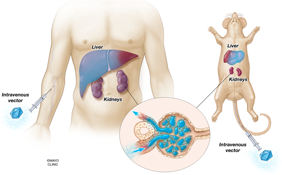
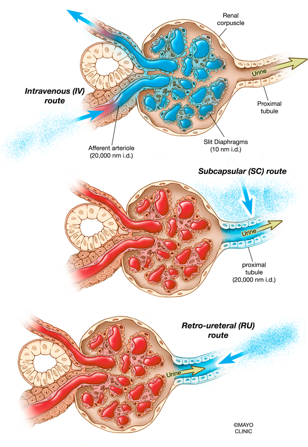
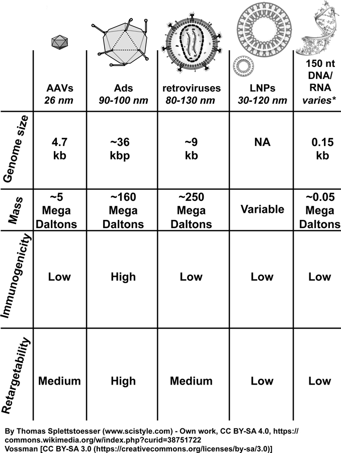
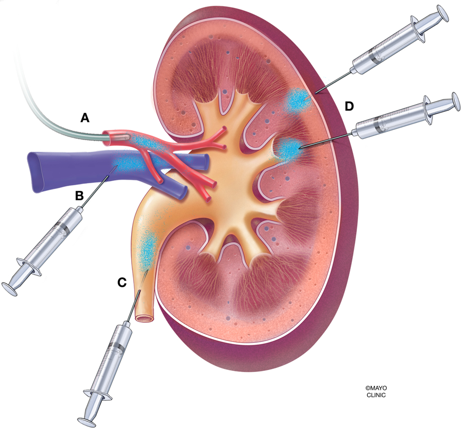
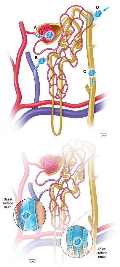
Mutations in approximately 80 genes have been implicated as the cause of various genetic kidney diseases. However, gene delivery to kidney cells from the blood is inefficient because of the natural filtering functions of the glomerulus, and research into and development of gene therapy directed toward kidney disease has lagged behind as compared with hepatic, neuromuscular, and ocular gene therapy. This lack of progress is in spite of numerous genetic mouse models of human disease available to the research community and many vectors in existence that can theoretically deliver genes to kidney cells with high efficiency. In the past decade, several groups have begun to develop novel injection techniques in mice, such as retrograde ureter, renal vein, and direct subcapsular injections to help resolve the issue of gene delivery to the kidney through the blood. In addition, the ability to retarget vectors specifically toward kidney cells has been underutilized but shows promise. This review discusses how recent advances in gene delivery to the kidney and the field of gene therapy can leverage the wealth of knowledge of kidney genetics to work toward developing gene therapy products for patients with kidney disease.


Figures

Figure 1.
Figure 1.. Expected target tissues for transduction after intravenous administration of typical viral vectors.
Liver-tropic vectors, such as AAV8 and Ad5, will highly transduce hepatocytes when administered systemically in large doses. Meanwhile, the vectors may physically reach the kidney from the blood, but will be limited to transducing cells in the glomerulus. Other tissues capable of being significantly transduced from systemic administration are the heart when AAV9 is used. Transduction is depicted as tissues and cells shaded in blue.
Figure 2.
Figure 2.. Intravenous and direct kidney vector administration in the context of the renal corpuscle.
Injecting vectors by the intravenous route would result in most vectors being stuck in the glomeruli of the kidneys or recirculated into the bloodstream, since slit diaphragms are approximately 10 nm in diameter (upper). Injecting vectors by the subcapsular route would bypass the glomeruli and potentially give vectors access to the apical and basolateral sides of some tubules (middle). Injecting vectors by the retro-ureteral route would theoretically grant vectors access to the apical side of tubules unless they escape from within the tubule epithelium, since proximal tubules are up to 20,000 nm in diameter (lower). nm, nanometer; i.d., in diameter.
Figure 3.
Figure 3.. Molecular therapy vectors and their respective sizes.
None of the molecular therapy vectors shown here, with the exception of 150 nt or smaller nucleic acids, would be expected to penetrate past the glomerular barrier and into the epithelium of the kidney, based on their respective masses and diameters. AAVs, adeno-associated viruses; Ads, adenoviruses; LNPs, lipid nanoparticles; nt, nucleotide; kb, kilobase; kbp, kilobasepair; kDa, kiloDalton.
Figure 4.
Figure 4.. Various modalities of vector administration in the context of the whole kidney.
(A) Infusion into the renal artery, via catheter or injection. (B) Retrograde infusion into the renal vein. (C) Retrograde infusion into the ureter. (D) Subcapsular injection into the parenchyma of the kidney
Figure 5.
Figure 5.. Routes of vector entry in the context of the nephron
(A) Blood enters the glomerulus of the nephron through the afferent arteriole, which has an approximate diameter of 20,000 nm. However, filtrate that penetrates further into the nephron is limited by the diameter of the slit diaphragms in the glomerular basement membrane, which are approximately 10 nm in diameter. The small diameter of the slit diaphragms excludes molecular therapy vectors from entering the kidney from the bloodstream, while the much broader diameter of the tubule epithelium (20,000 nm or greater) may allow entrance of vectors from the ureteral route. A high pressure delivery, I.E., a hydrodynamic injection, may change the principles of which vectors can pass through the glomerular barrier. (B) Vector access to the nephron after injection into the renal vein. Vectors may escape from the vasculature endothelium and access the basolateral surface of tubules (inset). (C) Vector access to the nephron after retrograde ureteral infusion. Vectors theoretically have exclusive access to the apical side of tubules, from the collecting duct to the proximal tubule (inset). (D) Vector access to the nephron after subcapsular injection. Vectors come into contact with tubules in a more undirected fashion, which theoretically is from “extra-tubular” space of kidney. Vectors may have access to basolateral or apical sides of tubules.

References

    1. Hill NR, Fatoba ST, Oke JL, Hirst JA, O’Callaghan CA, Lasserson DS, et al. Global Prevalence of Chronic Kidney Disease - A Systematic Review and Meta-Analysis. PLoS One. 2016;11(7):e0158765. - PMC - PubMed
    1. Hildebrandt F Genetic kidney diseases. Lancet. 2010. April 10;375(9722):1287–95. - PMC - PubMed
    1. Leung JC. Inherited renal diseases. Curr Pediatr Rev. 2014;10(2):95–100. - PubMed
    1. Boron WB, Emile. Glomerular Filtration and Renal Blood Flow Medical Physiology. 3rd ed: Elsevier; 2017. p. 739–53.e2.
    1. Murphy JJ, Myint MK, Rattner WH, Klaus R, Shallow J. The lymphatic system of the kidney. J Urol. 1958. July;80(1):1–6. - PubMed

Publication types