Review
. 2020 Oct 14;120(19):10695-10743.
doi: 10.1021/acs.chemrev.9b00810. Epub 2020 Apr 23.

Photopolymerizable Biomaterials and Light-Based 3D Printing Strategies for Biomedical Applications

Affiliations
Review

Photopolymerizable Biomaterials and Light-Based 3D Printing Strategies for Biomedical Applications

Claire Yu et al. Chem Rev. .

Abstract

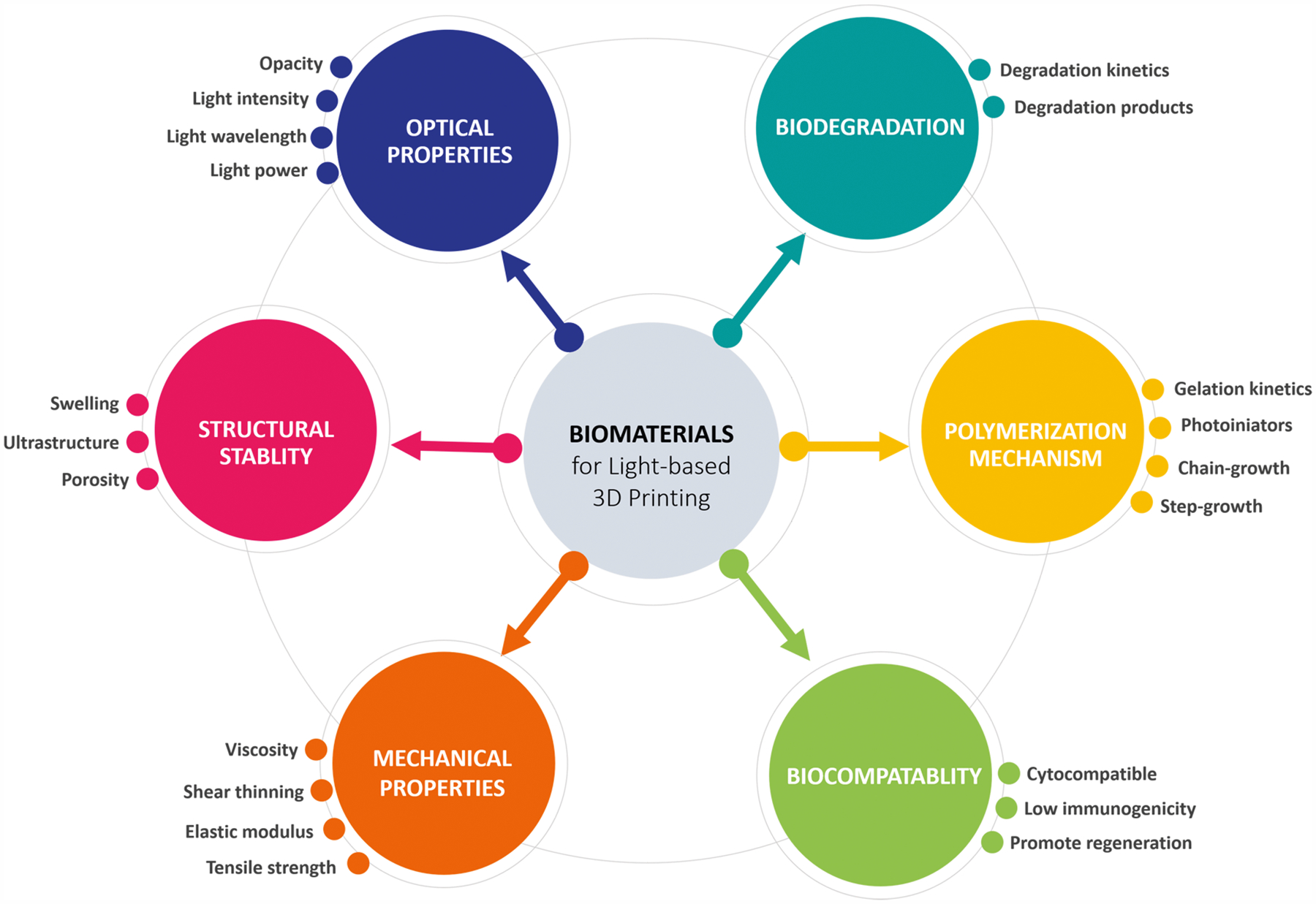
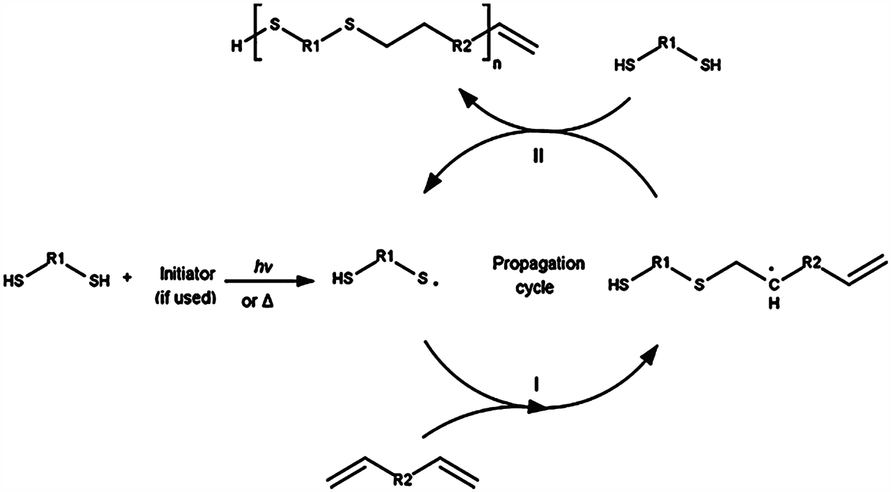
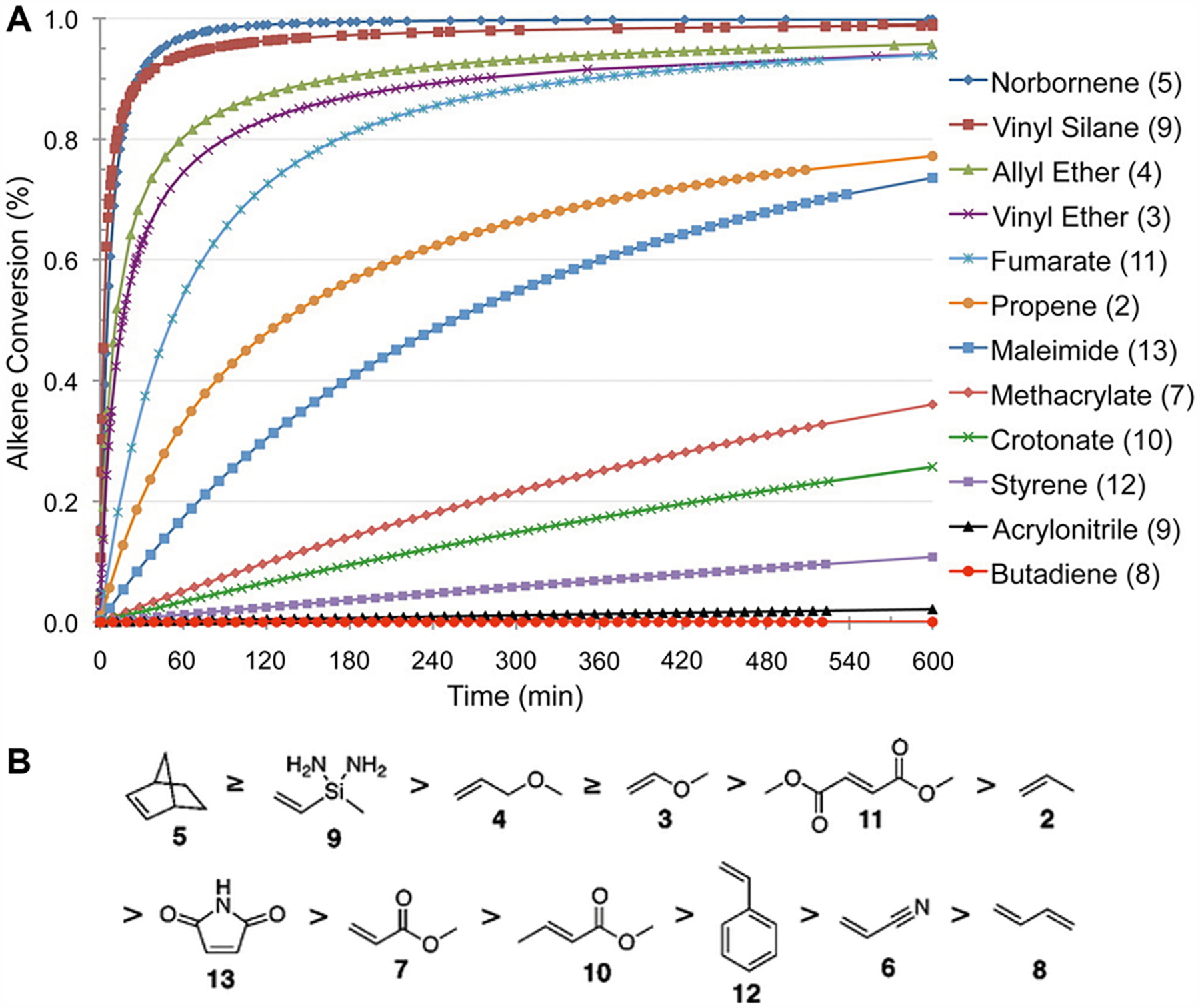
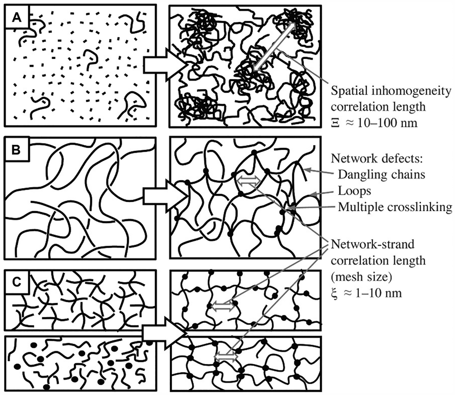
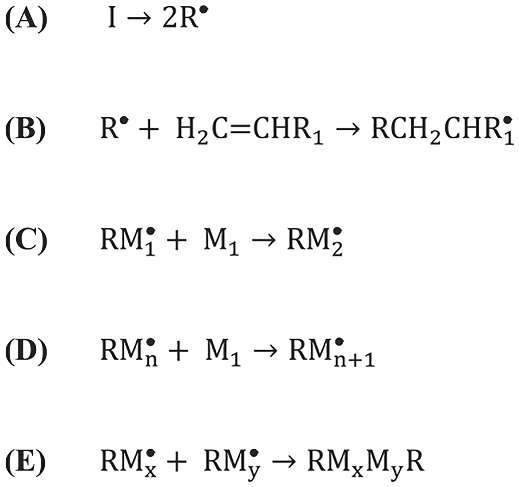
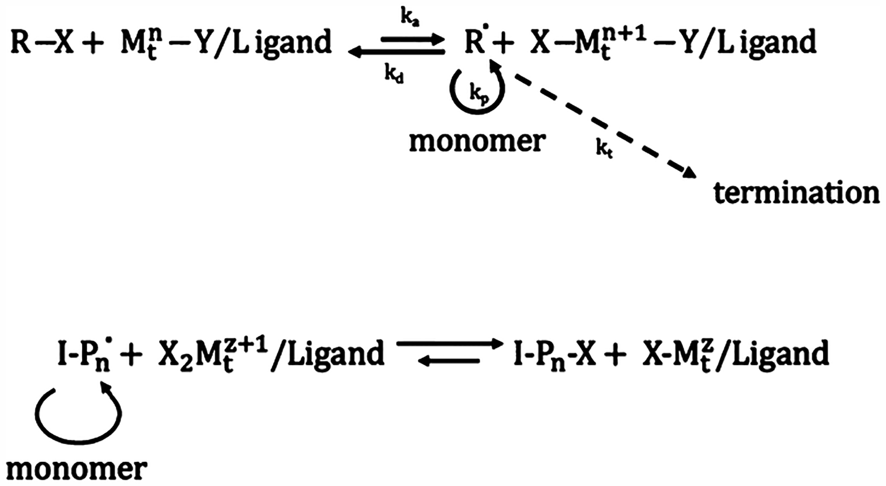
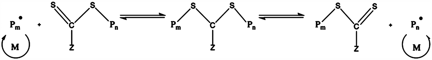
Since the advent of additive manufacturing, known commonly as 3D printing, this technology has revolutionized the biofabrication landscape and driven numerous pivotal advancements in tissue engineering and regenerative medicine. Many 3D printing methods were developed in short course after Charles Hull first introduced the power of stereolithography to the world. However, materials development was not met with the same enthusiasm and remained the bottleneck in the field for some time. Only in the past decade has there been deliberate development to expand the materials toolbox for 3D printing applications to meet the true potential of 3D printing technologies. Herein, we review the development of biomaterials suited for light-based 3D printing modalities with an emphasis on bioprinting applications. We discuss the chemical mechanisms that govern photopolymerization and highlight the application of natural, synthetic, and composite biomaterials as 3D printed hydrogels. Because the quality of a 3D printed construct is highly dependent on both the material properties and processing technique, we included a final section on the theoretical and practical aspects behind light-based 3D printing as well as ways to employ that knowledge to troubleshoot and standardize the optimization of printing parameters.


Conflict of interest statement

The authors declare no competing financial interest.

Figures

Figure 1.
Figure 1.
Overview of biomaterials selection criteria for light-based 3D printing in tissue engineering and regenerative medicine applications.
Figure 2.
Figure 2.
Free radical initiated thiol–ene click chemistry reaction mechanism. Propagation occurs in mechanism I. The initiator free radical abstracts the thiol hydrogen, producing a thiyl radical that attacks the alkene double bond. Chain transfer occurs in mechanism II. The thiyl radical is regenerated by the alkyl radical abstracting a free thiol hydrogen, which under the right reaction conditions will occur much more often than attacking another alkene double bond. The thiyl radical can now continue to propagate the thiol–ene reaction. Reproduced with permission from ref . Copyright 2017 Elsevier.
Figure 3.
Figure 3.
Effect of alkene group selection on thiol–ene reaction kinetics. (A) Theoretical computation of the kinetics of the thiol–ene reaction dependent on the reactivity of the chosen alkene group. Norbornene is a popular alkene candidate for thiol–ene reactions due to its superior reaction rate. Methacrylate, the common reactive group for chain-growth photopolymerization, has a starkly slow thiol–ene kinetics, with the alkene conversion well below 50% even after a 10 h reaction time. (B) Descending list of alkene group reactivity based on the theoretical kinetics model. Reprinted with permission from ref . Copyright 2012 American Chemical Society.
Figure 4.
Figure 4.
Depiction of hydrogel network formation depending on cross-linking mechanism and the resulting degree of inhomogeneity. (A) Free-radical chain growth polymerization of monomers and cross-linkers leading to spatial inhomogeneity within the network architecture. (B) Network formation via cross-linking of reactive functional side groups of the polymer chains in a semidilute solution, leading to local inhomogeneity. (C) Orthogonal step-growth polymerization resulting in a mostly ordered, homogeneous network. Reproduced with permission from ref . Copyright 2017 Elsevier.
Figure 5.
Figure 5.
Photolysis mechanism of o-nitrobenzyl (R1 = H) and nitrophenylethyl (R1 = methyl).
Figure 6.
Figure 6.
Various tissue constructs bioprinted with naturally derived biomaterials. (A) Schematic and bright-field image of a cantilever cardiac tissue model bioprinted with GelMA for measuring the cardiac contraction force. Scale bar: 500 μm. Reproduced with permission from ref . Copyright 2019 Elsevier. (B) Fluorescence and bright field images of a biomimetic multicellular liver tissue model bioprinted with GelMA and GM-HA for drug testing. Scale bars: 500 μm. Reproduced with permission from ref . Copyright 2016 National Academy of Sciences. (C) Digital designs and bright field images of biomimetic heart and liver tissues bioprinted with tissue-specific dECM bioinks. Scale bar: 1 mm. Reproduced with permission from ref . Copyright 2019 Elsevier. (D) Fluorescence and bright field images of a hepatic cancer model bioprinted with liver dECM bioink to recapitulate various stages of fibrotic liver disease. Scale bars: 500 μm. Reproduced with permission from ref . Copyright 2018 Elsevier.
Figure 7.
Figure 7.
Various 3D printed PEG-based hydrogel structures for cell biology. (A) 3D printed PEGDA patterns (from left to right: stripes, symmetric forks, and asymmetric forks) for investigating the impact of cellular alignment and stress on ADSC differentiation. Scale bars: 100 μm. (B) Immunofluorescent staining of smooth muscle α-actin revealing the cell alignment and myogenesis on the three PEGDA patterns. (A,B) Reproduced with permission from ref . Copyright 2013 Elsevier. (C) 3D printed microwells with various shapes for multicellular spheroid and embryoid body culture. Reproduced with permission from ref . Copyright 2012 Wiley-VCH. (D) Nature-inspired fractal patterns for investigating cell organization behaviors. Reproduced with permission from ref . Copyright 2016 American Chemical Society. (E) 3D printed web structures with microscale units featuring positive and negative Poisson’s ratios. Reproduced with permission from ref . Copyright 2013 Wiley-VCH.
Figure 8.
Figure 8.
Various 3D printed PEG-based hydrogel structures for tissue engineering and regenerative medicine. (A) 3D printed biomimetic spinal cord scaffold with microchannels for complete rat spinal cord transection. (B) 3D printed spinal cord scaffold based on MRI of human spinal cord injury. (A,B) Reprinted with permission from ref . Copyright 2019 Springer Nature. (C) Various 3D printed nerve guidance conduits (NGCs) for peripheral nerve regeneration. (D) 3D printed human life-size facial NGC. (C,D) Reproduced with permission from ref . Copyright 2018 Elsevier.
Figure 9.
Figure 9.
3D printed Nor-PGS as (A) open-lattice cube, (B) nose, and (C) ear shaped structures. Reproduced with permission from ref . Copyright 2017 Royal Society of Chemistry.
Figure 10.
Figure 10.
Polymerization mechanism of polyurethanes. (A) One-stage polymerization where polyols/polyamines and chain extenders react with excess diisocyanates simultaneously. (B) Two-stage polymerization where polyols/polyamines react with diisocyanates first, followed by an additional reaction with the chain extenders.
Figure 11.
Figure 11.
Common diisocyanates used in large-scale polyurethane productions.
Figure 12.
Figure 12.
Common oligodiols used in polyurethane production, including polyether, polyester, and polycarbonate-based oligodiols. The nature of oligodiols used will determine the properties of polyurethane synthesized.
Figure 13.
Figure 13.
Schematic drawings explaining the difference in polymer chain structures between thermoplastic and thermosetting polyurethanes. Thermoplastic polyurethanes will have higher backbone flexibilities, whereas thermosetting polyurethanes are generally more rigid. Reproduced with permission from ref . Copyright 2015 Multidisciplinary Digital Publishing Institute (MDPI).
Figure 14.
Figure 14.
Hard and soft segment distribution in PU. Reproduced with permission from ref . Copyright 2011 Elsevier.
Figure 15.
Figure 15.
Schematic of different types of nanomaterials that can be used to form nanocomposite hydrogels.
Figure 16.
Figure 16.
(A) Optical images of CNT/GelMA prepolymer solutions showing increasing optical density with increasing CNT concentration. (B) High resolution transmission electron microscopy image of well-dispersed 0.5 mg/mL CNT/GelMA prepolymer solution. (C) UV–vis adsorption spectra of prepolymer solutions. Absorption at 365 nm increases with increasing CNT concentration. (D) Fluorescence images of micropatterned CNT/GelMA hydrogels. CNTs functionalized with FITC for visualization. Scale bar: 300 μm. Reproduced with permission from ref . Copyright 2012 American Chemical Society.
Figure 17.
Figure 17.
3D printed microfish. (A) Energy-dispersive X-ray spectroscopy showing 3D microfish with different nanoparticles localized at the head, tail, and body. (B) Fluorescent image of the microfish after detoxification of a melittin solution. (C) Time-lapse images of the microfish performing sharp turns with magnetic guidance. (A–C) Reproduced with permission from ref . Copyright 2015 Wiley-VCH.
Figure 18.
Figure 18.
(A) Schematic of the mechanism of hydroxyapatite (HA) formation in the GelMA network. (B) Schematic of printing setup. HUVECs encapsulated in the prepolymer system were first micropatterned, followed by MG63 cells encapsulated into the prepolymer system. The printed rings are then assembled in a modular fashion into tubes. (C) Characterization of osteon-like double-ring modules. Phase-contrast images of micropatterned print of single unit as well as a full tube assembly. (D) Confocal image of cells in the structure at day 7. (E) Fluorescent image of the tube under rhodamine (red) perfusion. (F) Schematic of the cortical bone used as inspiration for print. Reproduced with permission from ref . Copyright 2015 American Chemical Society.
Figure 19.
Figure 19.
3D printed liver detoxification device. (A) Fluorescent image of 3D printed liver-inspired detoxification device with polydiacetylene nanoparticles encapsulated in PEGDA. (B) Scanning electron microscope image of this detoxification device. Scale bar: 50 mm. (C) The liver-inspired detoxification device demonstrated higher neutralization efficiency than the slab control. (A–C) Reproduced with permission from ref . Copyright 2014 Springer Nature.
Figure 20.
Figure 20.
Classification of light-based 3D printing modalities. (A) Primary configuration involves serial deposition of biomaterials in dot-by-dot or line-by-line fashion. (B) Secondary configuration involves planar build via digital light processing (DLP)-based projection of patterns into a biomaterial vat. (C) Tertiary configuration involves volumetric build via DLP-based projection of patterns into a rotating biomaterial vat.
Scheme 1.
Scheme 1.
General Initiation (A,B), Propagation (C,D), and Termination (E) Chemical Reactions for Free Radical Polymerization
Scheme 2.
Scheme 2.
(top) Generalized ATRP Reaction Mechanism; (bottom) Generalized Reverse-ATRP Reaction Mechanism
Scheme 3.
Scheme 3.
Generalized RAFT Reaction Mechanism

References

    1. Edmondson R; Broglie JJ; Adcock AF; Yang L Three-Dimensional Cell Culture Systems and Their Applications in Drug Discovery and Cell-Based Biosensors. Assay Drug Dev. Technol 2014, 12, 207–218. - PMC - PubMed
    1. Antoni D; Burckel H; Josset E; Noel G Three-Dimensional Cell Culture: A Breakthrough in Vivo. Int. J. Mol. Sci 2015, 16, 5517–5527. - PMC - PubMed
    1. Tibbitt MW; Anseth KS Hydrogels as Extracellular Matrix Mimics for 3D Cell Culture. Biotechnol. Bioeng 2009, 103, 655–663. - PMC - PubMed
    1. Frantz C; Stewart KM; Weaver VM The Extracellular Matrix at a Glance. J. Cell Sci 2010, 123, 4195–4200. - PMC - PubMed
    1. Ozbolat IT; Peng W; Ozbolat V Application Areas of 3D Bioprinting. Drug Discovery Today 2016, 21, 1257–1271. - PubMed

Publication types

Substances