. 2020 Aug;9(15):e1901792.
doi: 10.1002/adhm.201901792. Epub 2020 Apr 23.

One-Step Photoactivation of a Dual-Functionalized Bioink as Cell Carrier and Cartilage-Binding Glue for Chondral Regeneration

Affiliations

One-Step Photoactivation of a Dual-Functionalized Bioink as Cell Carrier and Cartilage-Binding Glue for Chondral Regeneration

Khoon S Lim et al. Adv Healthc Mater. 2020 Aug.

Abstract

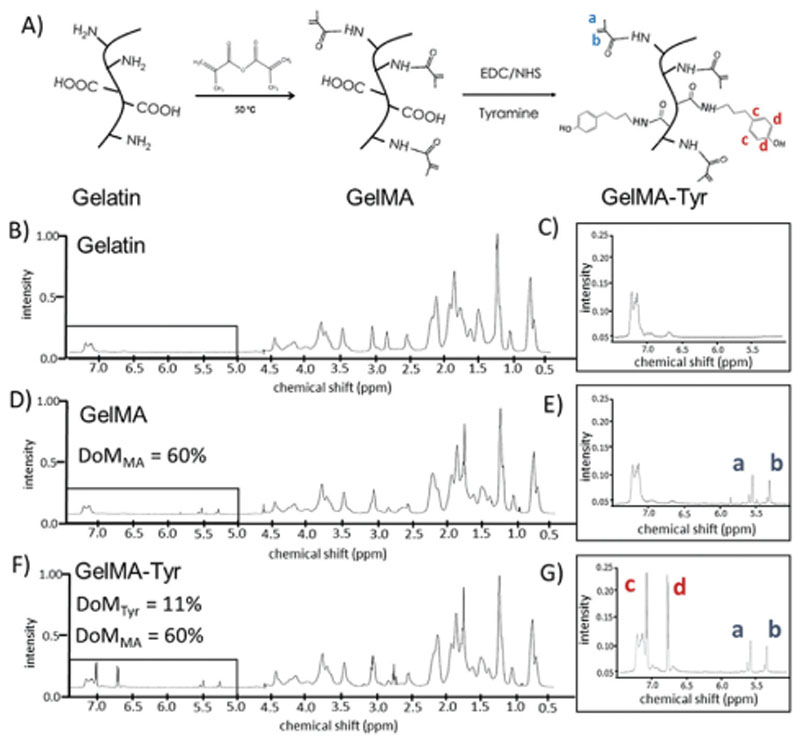
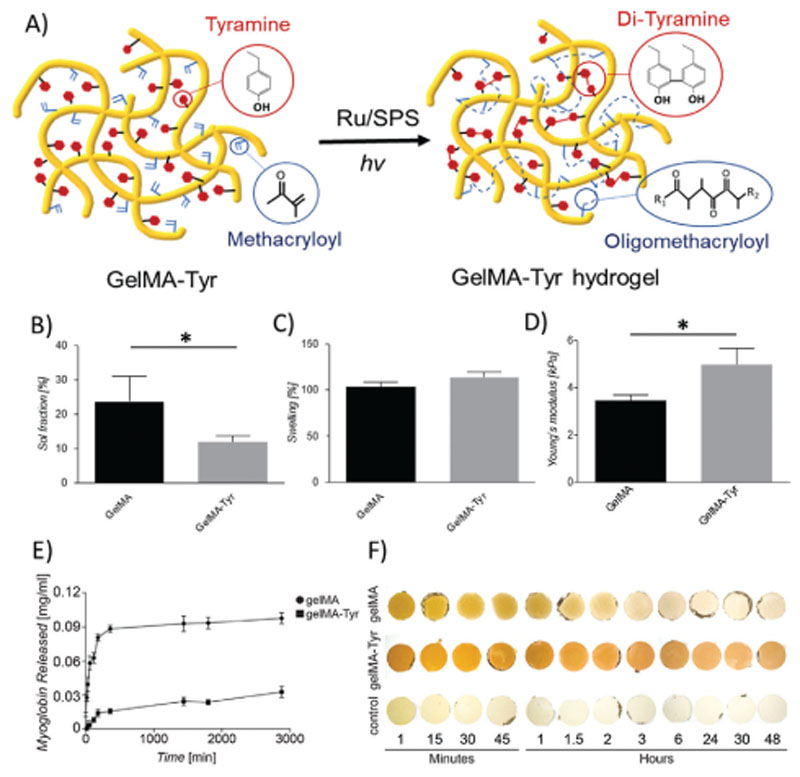
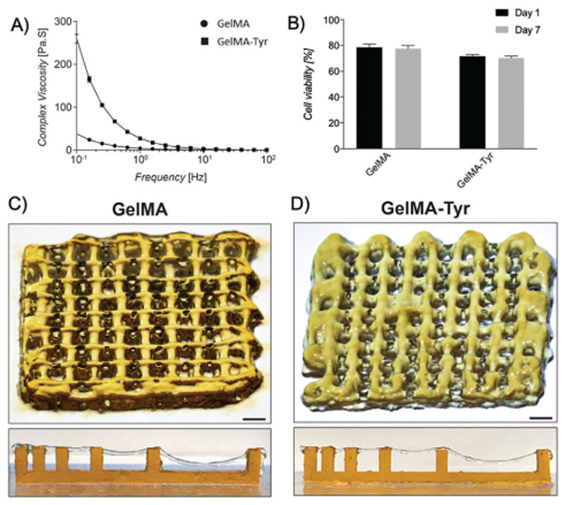
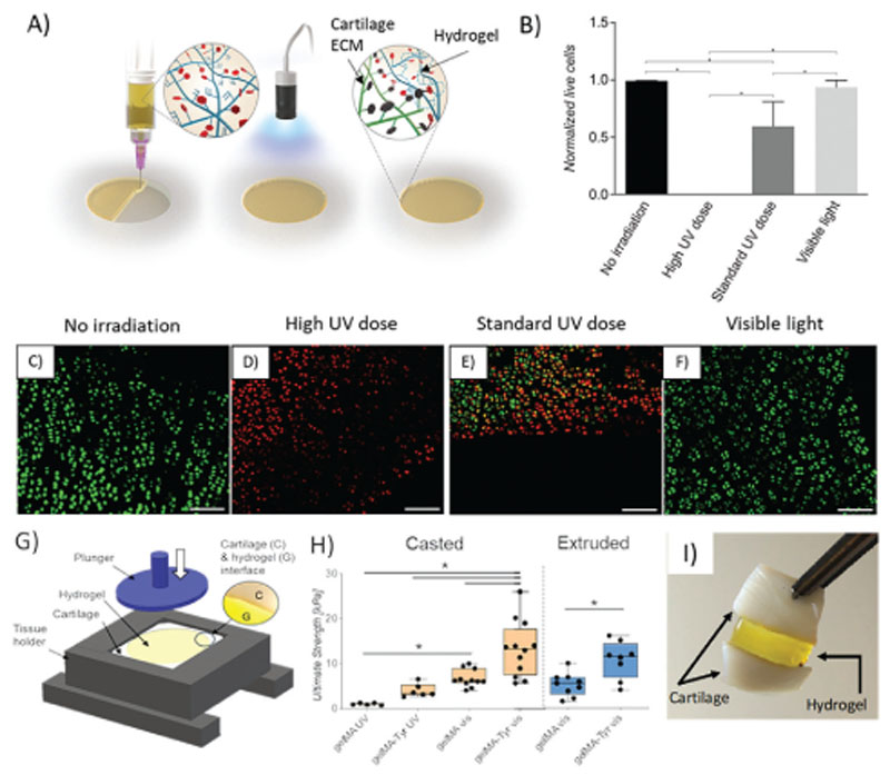
Cartilage defects can result in pain, disability, and osteoarthritis. Hydrogels providing a chondroregeneration-permissive environment are often mechanically weak and display poor lateral integration into the surrounding cartilage. This study develops a visible-light responsive gelatin ink with enhanced interactions with the native tissue, and potential for intraoperative bioprinting. A dual-functionalized tyramine and methacryloyl gelatin (GelMA-Tyr) is synthesized. Photo-crosslinking of both groups is triggered in a single photoexposure by cell-compatible visible light in presence of tris(2,2'-bipyridyl)dichlororuthenium(II) and sodium persulfate as initiators. Neo-cartilage formation from embedded chondroprogenitor cells is demonstrated in vitro, and the hydrogel is successfully applied as bioink for extrusion-printing. Visible light in situ crosslinking in cartilage defects results in no damage to the surrounding tissue, in contrast to the native chondrocyte death caused by UV light (365-400 nm range), commonly used in biofabrication. Tyramine-binding to proteins in native cartilage leads to a 15-fold increment in the adhesive strength of the bioglue compared to pristine GelMA. Enhanced adhesion is observed also when the ink is extruded as printable filaments into the defect. Visible-light reactive GelMA-Tyr bioinks can act as orthobiologic carriers for in situ cartilage repair, providing a permissive environment for chondrogenesis, and establishing safe lateral integration into chondral defects.

Keywords: biofabrication; bioglue; bioprinting; cartilage tissue engineering; tissue integration.


Conflict of interest statement

Conflict of Interest

The authors declare no conflict of interest.

Figures

Figure 1
Figure 1. Functionalization process to obtain the dual responsive hydrogels.
A) Schematic representation of the synthesis of GelMA and GelMA-Tyr; representative 1H-NMR spectra of gelatin, GelMA and GelMA-Tyr. B–G) 1H-NMR spectra of the functionalized hydrogels, showing the presence of the characteristic peaks for acrylates (a,b) in the GelMA and GelMA-Tyr groups and for the added phenolic groups in the GelMA-Tyr polymers.
Figure 2
Figure 2. Fabrication and physicochemical and mechanical characterization of the one-step inducible, dual crosslinked hydrogels.
A) Schematic representation of the crosslinking process of the GelMA-Tyr macromer. B) Sol fraction values and C) swelling behavior of the hydrogels as observed via sol–gel analysis; D) compressive young’s modulus of the casted hydrogels. Incorporation of myoglobin into GelMA or GelMA-Tyr hydrogels: E) release profile of myoglobin from GelMA or GelMA-Tyr hydrogels over 3000 min; F) macroscopic images of GelMA and GelMA-Tyr hydrogels incorporated with myoglobin over 48 h.
Figure 3
Figure 3. Chondrogenic differentiation of ACPCs encapsulated within GelMA or GelMA-Tyr hydrogels.
A) Cell viability after 1 and 7 days in culture. B) GAG/DNA and C) Young’s modulus of ACPC-laden GelMA or GelMA-Tyr gels after 1 and 28 days in culture. D) Collagen type II, E) Collagen type I, and F) PRG4 gene expression of ACPC-laden GelMA or GelMA-Tyr gels after 1 and 28 days in culture. Histological stainings of ACPC-laden GelMA or GelMA-Tyr gels after 28 days, G,J) safranin-O, H,K) collagen type II, scale bar = 20 μm, and I,L) collagen type 1, scale bar = 40 μm.
Figure 4
Figure 4. Extrusion bioprinting of GelMA-Tyr bioinks.
A) Rheological profile of GelMA and GelMA-Tyr in response to shear rate. B) Cell viability of ACPCs bioprinted within GelMA and GelMA-Tyr constructs. Macroscopic images of extrusion bioprinted C) GelMA and D) GelMA-Tyr constructs, together with a representative image of the filament collapse assay, showing the ability of the printed struts to bridge gaps of 4 mm without noticeable deformation, and up to 16 mm while experiencing sagging. Scale bar = 1 mm.
Figure 5
Figure 5
A) Schematic of intraoperative administration of GelMA-Tyr to the chondral defect. B) Normalized amount of live cells and C–F) LIVE/DEAD images of cartilage biopsies irradiated with UV or visible light. Scale bar = 100 μm. G) Setup of the pushout assay to determine bond-strength. H) Bond-strength of GelMA or GelMA-Tyr administered to the cartilage biopsies as a solution or physically crosslinked gel. I) Cartilage biopsies adhered together using GelMA-Tyr.

References

    1. Flanigan DC, Harris JD, Trinh TQ, Siston RA, Brophy RH. Med Sci Sports Exercise. 2010;42:1795. - PubMed
    1. Wluka AE, Ding C, Jones G, Cicuttini FM. Rheumatology. 2005;44:1311. - PubMed
    2. Mouser VHM, Dautzenberg NMM, Levato R, van Rijen MHP, Dhert W, Malda J, Gawlitta D. ALTEX. 2018;35:65. - PMC - PubMed
    1. Hunziker EB. Osteoarthritis Cartilage. 2002;10:432. - PubMed
    1. Vega SL, Kwon MY, Burdick JA. Eur Cells Mater. 2017;33:59. - PMC - PubMed
    1. Li J, Chen G, Xu X, Abdou P, Jiang Q, Shi D, Gu Z. Regener Bio-mater. 2019;6:129. - PMC - PubMed

Publication types