Review
. 2020 Apr 24;126(9):1260-1280.
doi: 10.1161/CIRCRESAHA.120.315937. Epub 2020 Apr 23.

Interleukin-1 and the Inflammasome as Therapeutic Targets in Cardiovascular Disease

Affiliations
Review

Interleukin-1 and the Inflammasome as Therapeutic Targets in Cardiovascular Disease

Antonio Abbate et al. Circ Res. .

Abstract

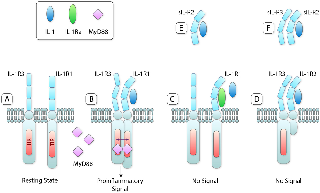
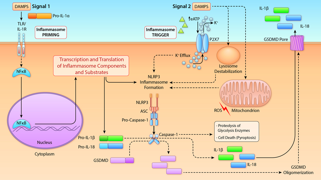
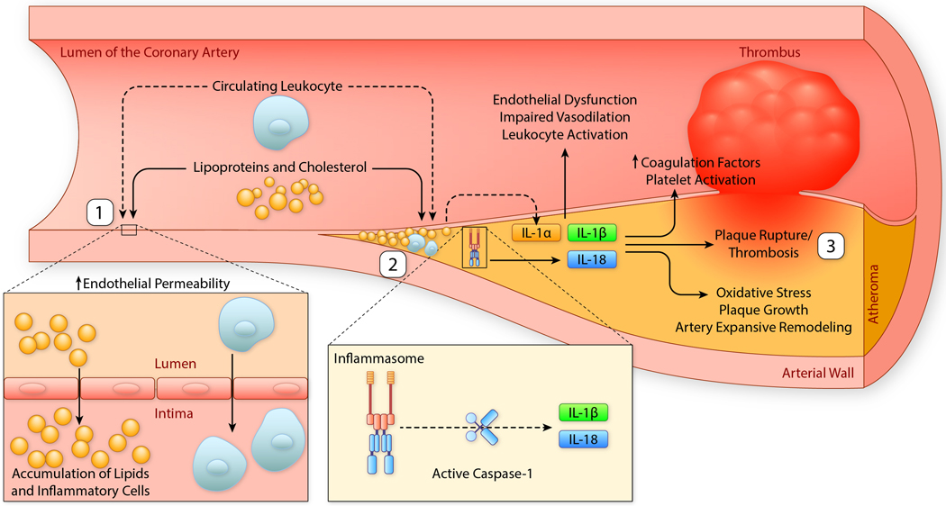
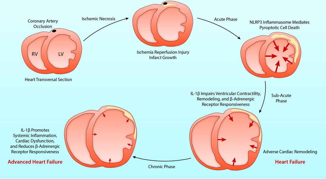
The intracellular sensing protein termed NLRP3 (for NACHT, LRR, and PYD domains-containing protein 3) forms a macromolecular structure called the NLRP3 inflammasome. The NLRP3 inflammasome plays a major role in inflammation, particularly in the production of IL (interleukin)-1β. IL-1β is the most studied of the IL-1 family of cytokines, including 11 members, among which are IL-1α and IL-18. Here, we summarize preclinical and clinical findings supporting the key pathogenetic role of the NLRP3 inflammasome and IL-1 cytokines in the formation, progression, and complications of atherosclerosis, in ischemic (acute myocardial infarction), and nonischemic injury to the myocardium (myocarditis) and the progression to heart failure. We also review the clinically available IL-1 inhibitors, although not currently approved for cardiovascular indications, and discuss other IL-1 inhibitors, not currently approved, as well as oral NLRP3 inflammasome inhibitors currently in clinical development. Canakinumab, IL-1β antibody, prevented the recurrence of ischemic events in patients with prior acute myocardial infarction in a large phase III clinical trial, including 10 061 patients world-wide. Phase II clinical trials show promising data with anakinra, recombinant IL-1 receptor antagonist, in patients with ST-segment-elevation acute myocardial infarction or heart failure with reduced ejection fraction. Anakinra also improved outcomes in patients with pericarditis, and it is now considered standard of care as second-line treatment for patients with recurrent/refractory pericarditis. Rilonacept, a soluble IL-1 receptor chimeric fusion protein neutralizing IL-1α and IL-1β, has also shown promising results in a phase II study in recurrent/refractory pericarditis. In conclusion, there is overwhelming evidence linking the NLRP3 inflammasome and the IL-1 cytokines with the pathogenesis of cardiovascular diseases. The future will likely include targeted inhibitors to block the IL-1 isoforms, and possibly oral NLRP3 inflammasome inhibitors, across a wide spectrum of cardiovascular diseases.

Keywords: atherosclerosis; cytokines; inflammasome; inflammation; interleukin.


Figures

Figure 1.
Figure 1.. IL-1 and IL-1 Receptors
A. Resting state of ligand binding chain of IL-1R1 and the co-receptor IL-1R3 on cell surfaces. B. Binding of IL-1β (or IL-1α not shown) to IL-1R1 results in a conformational change allowing IL-1R3 to bind resulting in a heterotrimeric complex. The TIR domains in the intracellular compartment approximate, MyD88 binds and triggers a pro-inflammatory signal via NFκB (not shown). C. IL-1Ra binds to IL-1R1 but does not cause a conformational change. IL-1R3 does not bind, a trimeric complex is not formed and there is no signal despite the presence of IL-1β. The affinity for IL-1Ra for IL-1R1 is greater than that for IL-1α and least for IL-1β. D. IL-1β binds to IL-1R2 and a conformational change occurs, IL-1R3 binds and there is a trimeric complex. Since IL-1R2 lacks an intracellular domain, there is no TIR domain to dimerize with the TIR domain on IL-1R3. There is no signal. E. soluble IL-1R2 in the extracellular space binds IL-1β and sequesters IL-1β away from IL-1R1 on cells. F. Soluble IL-1R2 binds IL-1β and forms a complex with soluble IL-1R3 resulting in a higher affinity for IL-1β. Abbreviations: IL-1, interleukin-1; IL-1R1, ligand binding IL-1 Receptor type 1; IL-1R3, co-receptor IL-1 Receptor type 3; IL-1Ra, IL-1 Receptor antagonist; MyD88, Myeloid Differentiation Factor 88 IL-1R2, IL-1 receptor type 2; sIL-1R2, soluble (extracellular domain) IL-1R2sIL-1R3, soluble IL-1R3; TIR, Toll-IL-1-Receptor domain. IIllustration credit: Ben Smith
Figure 2.
Figure 2.. Schematic of the NLRP3 inflammasome formation.
In most cells, including macrophages resident cells, activation of the NLRP3 inflammasome activation requires two signals. Signal 1 (left) is a priming step. Tissue damage, such as ischemia reperfusion injury, promotes the release of DAMPs, including the IL-1α precursor (pro-IL-1α) and eATP. The Signal 1 priming is initiated when DAMPs activate membrane receptors, including the TLRs or the IL-1R1, leading to the translocation of NF-κB into the nucleus. This event promotes the transcription and translation of several of pro-inflammatory genes, particularly the precursors of IL-1β and IL-18, as well as components of the NLRP3 inflammasome. IL-1β and IL-18 precursors accumulate in the cytosol and signal 1 does not directly result in NLRP3 activation. Signal 2 provides the trigger for activation of the inflammasome. This signal is promoted by eATP or intracellular DAMPs (e.g. mitochondrial molecules, like ROS, or lysosomal content) which in most cases involve the efflux of K+. In the heart, eATP activates the purinergic receptor P2X7, resulting in K+ efflux with subsequent activation of NLRP3. NLRP3 oligomerizes and binds ASC and pro-caspase-1 resulting in the auto-catalytic cleavage of pro-caspase-1 to active caspase-1. Caspase-1 cleaves pro-IL-1β and pro-IL-18 into their mature and active forms. Caspase-1 also cleaves gasdermind D (GSDMD) producing an N-terminal fragment that oligomerizes and forms plasma membrane pores. These pores facilitate the release of mature IL-1β and IL-18 into the extracellular space. Active Caspase-1 induces degradation of glycolysis enzymes thus reducing the energy production in the cell. Caspase-1 and GSDMD pores also mediate a form of regulated cell death termed pyroptosis. IIllustration credit: Ben Smith Abbreviations: ASC, apoptosis-associated spec-like protein containing a carboxy-terminal containing a caspase recruiting domain; DAMPs, damage associated molecular patterns; eATP, extracellular adenosine triphosphate; GSDMD, gasdermin D; IL-1α, interleukin-1α; IL-1β, interleukin-1β; IL-18, interleukin-18; IL-1R, Interleukin-1 receptor type 1; NF-κB, Nuclear factor-κB; NLRP3, NACHT LRR and PYD domains-containing protein 3; P2X7, purinergic receptor 2X7; ROS, reactive oxygen species; TLR, Toll-Like Receptors.
Figure 3.
Figure 3.. IL-1, inflammasome and atherothrombosis
Atherosclerosis is a chronic process that can culminate with plaque rupture and atherothrombosis. (1) Pro-inflammatory lipoproteins in the lumen of the arteries promote endothelial dysfunction and permeability, leading to accumulations of lipids and migration of pro-inflammatory cells (leukocytes) in the intima of the vessel. (2) Over time, the accumulation of lipids, including cholesterol, and inflammatory cells leads to production of cytokines, including IL-1α, NLRP3 inflammasome activation, and production of inflammasome dependent cytokines IL-1β and IL-18. This process perpetuates endothelial dysfunction, impairs vasodilation, activates leukocytes and promotes oxidative stress, plaque growth and arterial expansive (outward) remodeling. (3) IL-1 activity increases coagulation factors, contributes to platelet activation and promotes plaque rupture and thrombosis. IIllustration credit: Ben Smith Abbreviations: IL-1α, interleukin-1α; IL-1β, interleukin-1β; IL-18, interleukin-18; NLRP3, NACHT, LRR, and PYD domains-containing protein 3.
Figure 4.
Figure 4.. IL-1 and the inflammasome in AMI and heart failure
Prolonged coronary artery occlusion leads to necrosis of the cardiomyocytes and acute myocardial infarction (AMI). In the acute phase, reperfusion limits necrosis but it does not interrupt the inflammatory response. Activation of the inflammasome following ischemia and reperfusion contributes to the acute loss of cardiomyocytes through pyroptotic cells death. In the subacute phase, locally produced IL-1β reduces myocardial contractility and the myocardial response to β-adrenergic receptor agonists, favoring adverse ventricular remodeling and heart failure. In the chronic phase, locally or systemically produced IL-1β perpetuates the contractile dysfunction, impaired β-adrenergic receptor responsiveness, progression of adverse ventricular remodeling and worsening heart failure. Cross-sections of the ventricles are depicted. The arrows represent the movement and force of the ventricular walls, with progressive weakening of contractility over time. IIllustration credit: Ben Smith Abbreviations: AMI, acute myocardial infarction; IL-1β, interleukin-1β; LV, left ventricle; RV, right ventricle.

References

    1. Dinarello CA. The IL-1 family of cytokines and receptors in rheumatic diseases. Nat Rev Rheumatol. 2019;15:612–632. - PubMed
    1. Dinarello CA. Interleukin-1 in the pathogenesis and treatment of inflammatory diseases. Blood. 2011;117:3720–32. - PMC - PubMed
    1. Van Tassell BW, Toldo S, Mezzaroma E and Abbate A. Targeting interleukin-1 in heart disease. Circulation. 2013;128:1910–23. - PMC - PubMed
    1. Buckley LF and Abbate A. Interleukin-1 blockade in cardiovascular diseases: a clinical update. Eur Heart J. 2018;39:2063–2069. - PubMed
    1. Buckley LF and Abbate A. Interleukin-1 Blockade in Cardiovascular Diseases: From Bench to Bedside. BioDrugs. 2018;32:111–118. - PubMed

MeSH terms