

The Multifaceted Nature of Tumor Microenvironment in Breast Carcinomas
- PMID: 32325459
- PMCID: PMC7265767
- DOI: 10.1159/000507055
The Multifaceted Nature of Tumor Microenvironment in Breast Carcinomas
Abstract
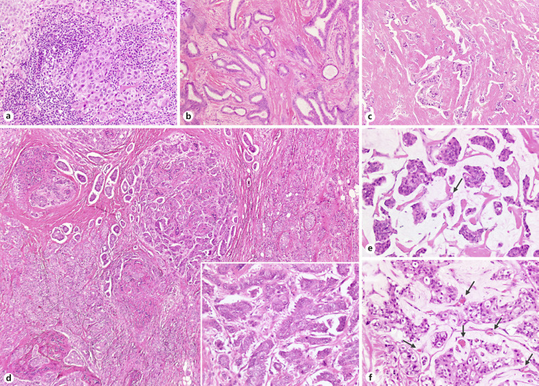
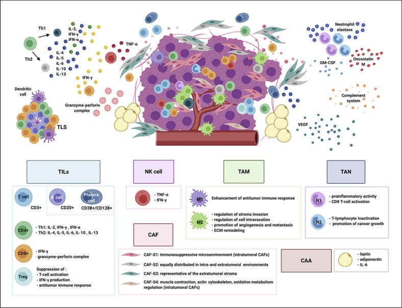
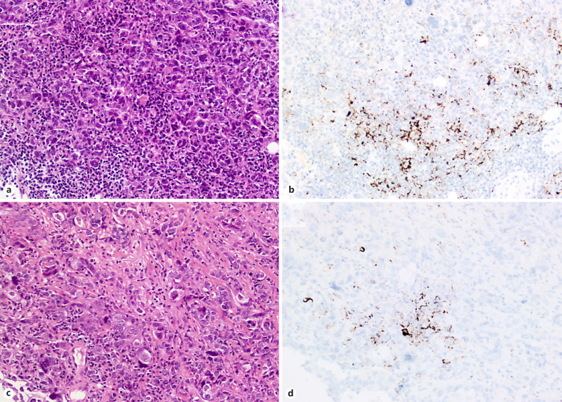
Heterogeneity in breast carcinomas can be appreciated at various levels, from morphology to molecular alterations, and there are well-known genotypic-phenotypic correlations. Clinical decision-making is strictly focused on the evaluation of tumor cells and is based on the assessment of hormone receptors and of the HER2 status, by means of a combination of immunohistochemical and in situ hybridization techniques. The tumor microenvironment (TME) also shows a multifaceted nature stemming from the different actors populating the intratumoral and the peritumoral stroma of breast carcinomas. Of note, we have now evidence that tumor-infiltrating lymphocytes (TILs) are clinically meaningful as their quantification in the intratumoral stroma strongly correlates with good prognosis, in particular in triple-negative and HER2-positive breast cancer patients. Nevertheless, TILs are just one of the many actors orchestrating the complexity of the TME, which is populated by immune and non-immune cells (cancer-associated fibroblasts, cancer-associated adipocytes), as well as non-cellular components such as chemical inflammation mediators. In this review article we will overview the main features of the distinct cell compartments by discussing (i) the potential impact the TME may have on the prognostic stratification of breast cancers and (ii) the possible predictive value of some markers in the context of immunotherapy in light of the recent results of phase III studies in advanced and early triple-negative breast cancer patients.
Keywords: Breast cancer; Cancer-associated fibroblasts; Immune cells; Immunotherapy; Ligand 1 of programmed cell death protein 1; Mutational load; Tumor-infiltrating lymphocytes.
© 2020 S. Karger AG, Basel.
Conflict of interest statement
C.M. has received personal/consultancy fees from Axiom Healthcare Strategies, Daiichi-Sankyo, MSD, Roche, Bayer, Thesaro, COR2ED. All of the other authors have no potential conflicts of interest to disclose.
Figures
References
-
- Lokuhetty D, White V, Watanabe R, Cree I. WHO Classification of Breast Tumours second volume in the 5th ed. 2019.
-
- Nakagawa S, Miki Y, Miyashita M, Hata S, Takahashi Y, Rai Y, et al. Tumor microenvironment in invasive lobular carcinoma: possible therapeutic targets. Breast Cancer Res Treat. 2016 Jan;155((1)):65–75. - PubMed
-
- Denkert C, von Minckwitz G, Darb-Esfahani S, Lederer B, Heppner BI, Weber KE, et al. Tumour-infiltrating lymphocytes and prognosis in different subtypes of breast cancer: a pooled analysis of 3771 patients treated with neoadjuvant therapy. Lancet Oncol. 2018 Jan;19((1)):40–50. - PubMed
-
- Marchiò C, Geyer F, Reis-Filho JS. Pathology and Molecular Pathology of Breast Cancer. Pathology and Epidemiology of Cancer; 2016. pp. pp.173–231.
-
- Dias AS, Almeida CR, Helguero LA, Duarte IF. Metabolic crosstalk in the breast cancer microenvironment. Eur J Cancer. 2019 Nov;121:154–71. - PubMed
Publication types
MeSH terms
Substances
LinkOut - more resources
Full Text Sources
Other Literature Sources
Medical
Research Materials
Miscellaneous
