. 2020 Apr 19;6(2):51.
doi: 10.3390/jof6020051.

Silver/Chitosan Nanocomposites: Preparation and Characterization and Their Fungicidal Activity against Dairy Cattle Toxicosis Penicillium expansum

Affiliations

Silver/Chitosan Nanocomposites: Preparation and Characterization and Their Fungicidal Activity against Dairy Cattle Toxicosis Penicillium expansum

Mousa A Alghuthaymi et al. J Fungi (Basel). .

Abstract

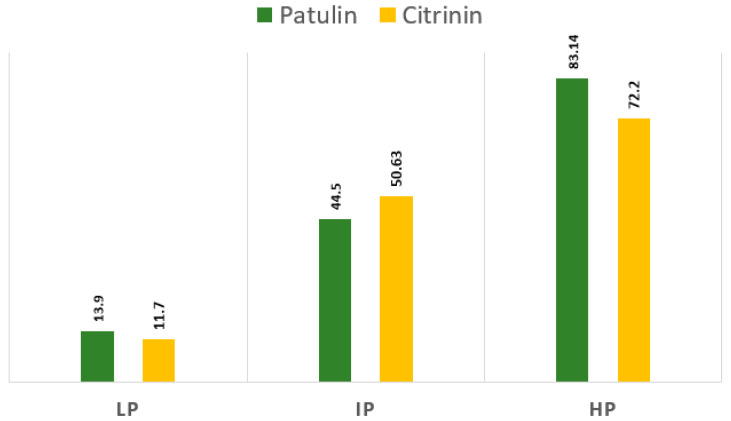
This work aimed to evaluate the fungicide activity of chitosan-silver nanocomposites (Ag-Chit-NCs) against Penicillium expansum from feed samples. The physicochemical properties of nanocomposites were characterized by X-ray fluorescence analysis (XRF), small-angle X-ray scattering (SAXS), X-ray photoelectron spectroscopy (XPS), and transmission electron microscopy (TEM). The morphological integrity of the nanohybrid was confirmed by electron transmission. By the data of RFA (X-ray fluorescent analysis), the contents of Ag in Ag-chitosan composite were 5.9 w/w%. The size distribution of the Ag nanoparticles incorporated in the chitosan matrix was investigated by SAXS. The main part of the size heterogeneity distribution in the chitosan matrix corresponds to the portion of small particles (3-4 nm). TEM analysis revealed a spherical morphology in the form of non-agglomerated caps, and 72% of the nanoparticles measured up to 4 nm. The minimum inhibitory concentration of NCs was evaluated in petri dishes. Three different concentrations were tested for antifungal activity against the mycotoxigenic P. expansum strain. Changes in the mycelium structure of P. expansum fungi by scanning electron microscopy (SEM) were observed to obtain information about the mode of action of Ag-Chit-NCs. It was shown that NC-Chit-NCs with sizes in the range from 4 to 10 nm have internalized sizes in cells, form agglomerates in the cytoplasm, and bind to cell organelles. Besides, their ability to influence protein and DNA fragmentation was examined in P. expansum. SDS-PAGE explains the apparent cellular protein response to the presence of various Ag-Chit-NCs. The intensity of P. expansum hyphal cell protein lines treated with Ag-Chit-NCs was very thin, indicating that high molecular weight proteins are largely prevented from entering the electrophoretic gel, which reflects cellular protein modification and possible damage caused by the binding of several protein fragments to Ag-Chit-NCs. The current results indicate that Ag-Chit-NCs <10 nm in size have significant antifungal activity against P. expansum, the causative agent of blue mold-contaminated dairy cattle feed.

Keywords: P. expansum; citrinin; mycotoxins; nanocomposites; patulin.


Conflict of interest statement

The authors declare no conflict of interest. The funders had a main role in the design of the study; in the collection, analyses, or interpretation of data; in the writing of the manuscript, or in the decision to publish the results.

Figures

Figure 1
Figure 1
Structural formula of patulin and citrinin (A) Patulin, (2,4-dihydroxy-2H-pyran-3 (6H) ylidene) acetic acid, (B) Citrinin, acid 3,4-lactone, (3R, 4S)-7-(Dihydroxymethylene))-3,4,5-trimethyl-3H-isochromic-6,8 (4H, 7H)-dione. Red dot (H) Blue dot (C). Data available online from: http://www.chemspider.com.
Figure 2
Figure 2
1—volume size distribution function of the scattering heterogeneities in initial high molecular weight chitosan after exposition in SC CO2 (25 MPa, 80 °C); 2—size distribution function D v (R) of the high molecular weight chitosan sample after impregnation with Ag-complex and reduction with hydrogen (1.15 MPa, 65 °C). Sample 1: chitosan (HMWC), Sample 2: chitosan matrix.
Figure 3
Figure 3
The C 1s spectra of samples 1 and 2 and their difference spectrum 3.
Figure 4
Figure 4
The Ag 3d and Ag MNN spectra of sample 1.
Figure 5
Figure 5
(A) Particle size analyser data, (B) Transmission electron microscopic image of Ag NPs Chitosan nanocomposites.
Figure 6
Figure 6
The quantitative results of patulin and citrinin assays of P. expansum isolates, including three levels of mycotoxins, high producer (HP), intermediate producer (IP), and low producer (LP).
Figure 7
Figure 7
Antifungal activity for different concentration of Ag-Chit-NCs (C1 = 0.30, C2 = 0.60, and C3 = 0.100 mg·mL−1) against P. expansum isolated from feeds by the plate assay. All Petri dish treatments were incubated at 28 °C for 10 days.
Figure 8
Figure 8
Protein expression profile of silver-stained SDS-PAGE from P. expansum mats at a high Ag-Chit-NCs concentration. Lane M contains a protein marker. Protein bands are shown in the black arrow (seven reduced band). The protein standards with molecular weights ranging from 66, 45, and 22 kDa were used. P. expansum isolates, including three levels of mycotoxins, high producer (HP), intermediate producer (IP), and low producer (LP). The same isolates treated with Ag-Chit NCs as a fellow T1, T2, and T3, respectively.
Figure 9
Figure 9
Agarose gel electrophoretic pattern of the fungal genomic DNA treated with 10 μL Ag-Chit-NCs. A: Lanes 1–3: DNA for untreated P. expansum isolates, Lanes 1: P. expansum 1, Lane 2: P. expansum 2, Lane 3: P. expansum 3. B: Lanes 1–6: three P. expansum isolates DNA treated with Ag-Chit nanocomposites (30, and 40 μg·mL−1) showed total damage to fragmented DNA bands.
Figure 10
Figure 10
Scanning electron microscope (SEM) photographs of P. expansum after treatment with Ag-Chit-NCs (a) P. expansum have globose smooth-walled and ellipsoidal conidia and are from 3 to 3.5 µm long (d). Red arrows refer to Ag-Chit NCs’ upper different fungal structures while blue arrows refer to the morphological changes in the fungal hyphae, such as irregular branching (b,c), loss of linearity and warty surfaces (c,f,h), collapsed cell, formation of a layer of extruded material (g,h,i), a small vesicle (i), SEM were the markedly shriveled, crinkled cell walls, and flattened hyphae of the fungi (gi), hyphal cell wall, and vesicle damage (ci).
Figure 11
Figure 11
The mechanistic approach of the antifungal action indicating ROS generation induced by Ag-Chit-NCs (star). Degradation and leakage of cell walls and cell membranes, electronic transport chain disturbances, enzyme inhibition, decomposition or destabilization of ribosomes, DNA and mRNA damage, and protein denaturation.

References

    1. Raper K.B., Thom C. A Manual of the Penicillia. Williams and Wilkins; Baltimore, MA, USA: 1949.
    1. Mansfield M.A., Jones A.D., Kuldau G.A. Contamination of fresh and ensiled maize by multiple Penicillium mycotoxins. Phytopathology. 2008;98:330–336. doi: 10.1094/PHYTO-98-3-0330. - DOI - PubMed
    1. Tangni E.K., Wambacq E., Bastiaanse H., Haesaert G., Pussemier L., De Poorter J., Foucart G., Van Hove F. Survey of fungal diversity in silages supplied to dairy cattle in Belgium over a two-year period. J. Anim. Sci. Adv. 2017;7:1861–1873.
    1. Pleadin J., Frece J., Markov K. Mycotoxins in food and feed. Adv. Food Nutr. Res. 2019;89:297–345. - PubMed
    1. Greco M., Pardo A., Pose G. Mycotoxigenic fungi and natural co-occurrence of mycotoxins in rainbow trout (Oncorhynchus mykiss) feeds. Toxins. 2015;7:4594–4609. doi: 10.3390/toxins7114595. - DOI - PMC - PubMed

LinkOut - more resources