Review
. 2020 Apr 20;21(8):2872.
doi: 10.3390/ijms21082872.

Non-Coding RNAs as Key Regulators of Glutaminolysis in Cancer

Affiliations
Review

Non-Coding RNAs as Key Regulators of Glutaminolysis in Cancer

Yunuen Ortiz-Pedraza et al. Int J Mol Sci. .

Abstract

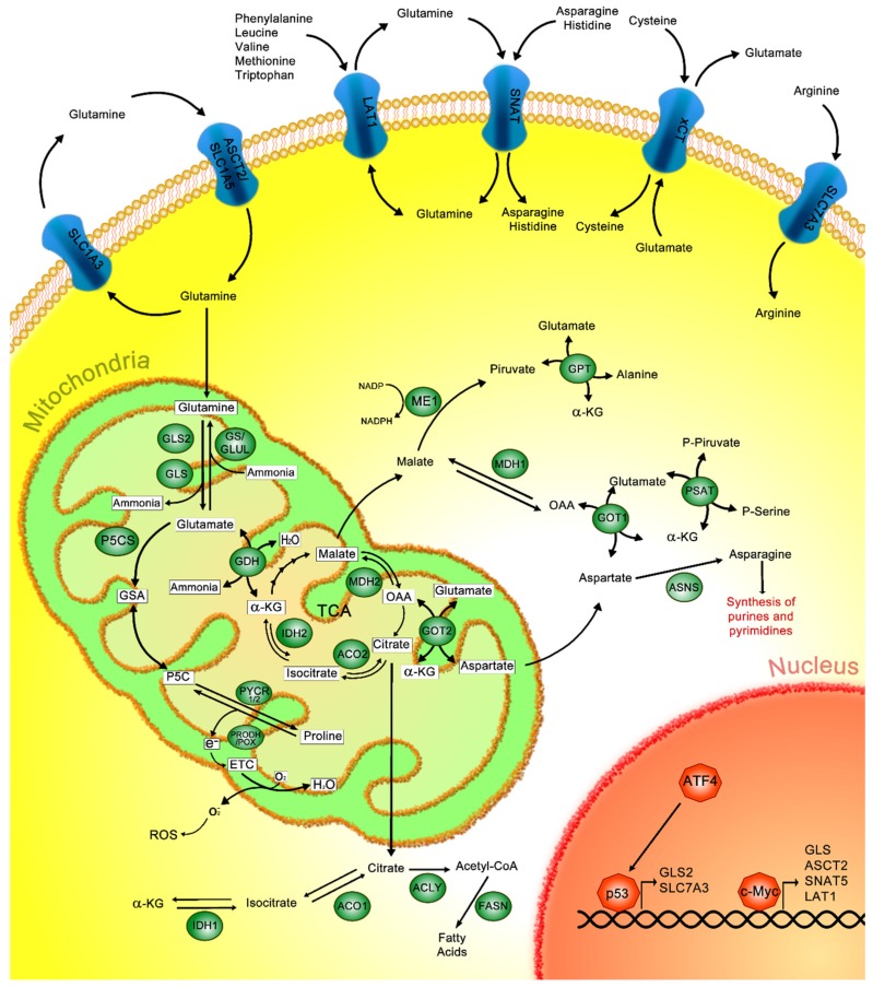
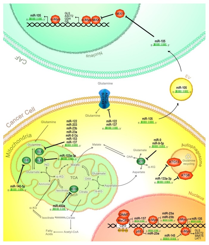
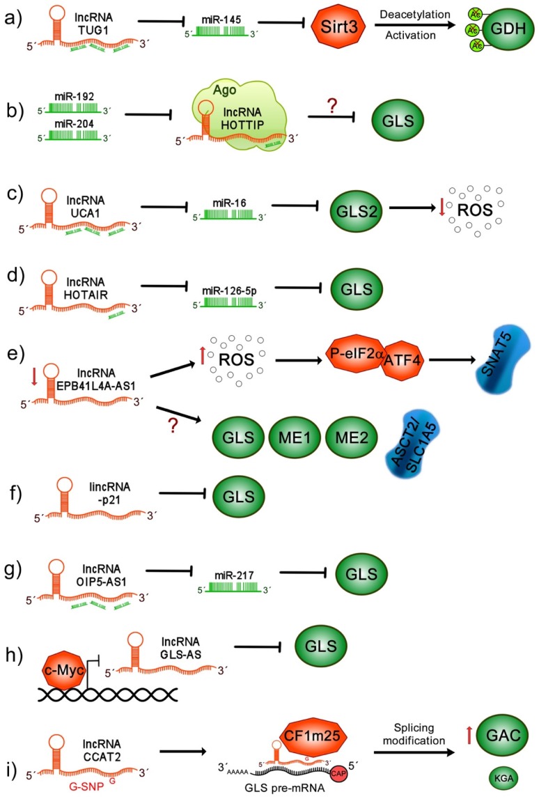
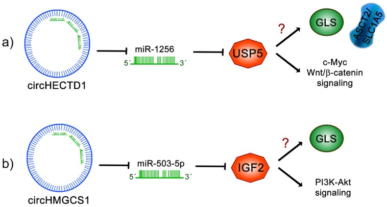
Cancer cells exhibit exacerbated metabolic activity to maintain their accelerated proliferation and microenvironmental adaptation in order to survive under nutrient-deficient conditions. Tumors display an increase in glycolysis, glutaminolysis and fatty acid biosynthesis, which provide their energy source. Glutamine is critical for fundamental cellular processes, where intermediate metabolites produced through glutaminolysis are necessary for the maintenance of mitochondrial metabolism. These include antioxidants to remove reactive oxygen species, and the generation of the nonessential amino acids, purines, pyrimidines and fatty acids required for cellular replication and the activation of cell signaling. Some cancer cells are highly dependent on glutamine consumption since its catabolism provides an anaplerotic pathway to feed the Krebs cycle. Intermediate members of the glutaminolysis pathway have been found to be deregulated in several types of cancers and have been proposed as therapeutic targets and prognostic biomarkers. This review summarizes the main players in the glutaminolysis pathway, how they have been found to be deregulated in cancer and their implications for cancer maintenance. Furthermore, non-coding RNAs are now recognized as new participants in the regulation of glutaminolysis; therefore, their involvement in glutamine metabolism in cancer is discussed in detail.

Keywords: cancer; glutaminolysis; lncRNAs; miRNAs.


Conflict of interest statement

The authors declare no conflict of interest.

Figures

Figure 1
Figure 1
Canonical glutaminolysis pathway. Glutamine is captured in the outer cell membrane through different amino acid transporters such as solute carrier (SLC)1A3, alanine, serine, cysteine, and glutamate transporter (ASCT2/SLC1A5), L-type amino acid transporter 1 (LAT1), sodium-neutral amino acid transporters (SNATs), sodium-independent cysteine–glutamate antiporter (xCT) or SLC7A3. Once in the mitochondria, glutamine is converted to glutamate through glutaminase 1 and glutaminase 2 (GLS, GLS2); inversely, glutamine synthetase (GS) can generate glutamine from glutamate. Glutamate dehydrogenase (GDH) catalyzes the conversion of glutamate to α-ketoglutarate (α-kG) and ammonia. Additionally, α-kG can also be obtained from the isocitrate that is derived from the tricarboxylic acid cycle (TCA); this reaction is catalyzed by isocitrate dehydrogenase 2 (IDH2), and isocitrate is previously obtained from citrate by the activity of aconitase 2 (ACO2). In the cytoplasm, isocitrate dehydrogenase 1 (IDH1) mediates the conversion of α-kG to isocitrate, then isocitrate is transformed into citrate through aconitase 1 (ACO1), which, in turn, is converted to acetyl-coenzyme A (acetyl-CoA) via adenosine triphosphate (ATP) citrate lyase (ACLY), finally producing fatty acids through the activity of fatty acid synthase (FASN). Moreover, α-kG is generated from oxaloacetate (OAA) by glutamate–oxaloacetate transaminases 1 and 2 (GOT1 and GOT2) in the cytoplasm and mitochondria, respectively. Then, cytoplasmic OAA is converted to malate by malate dehydrogenase 1 (MDH1), and further to pyruvate and nicotinamide adenine dinucleotide phosphate (NADPH) by malic enzyme 1 (ME1). Meanwhile, mitochondrial OAA is formed from malate by malate dehydrogenase 2 (MDH2). Additionally, glutamate is transformed to glutamic-γ-semialdehyde (GSA) by delta-1-pyrroline-5-carboxylate synthase (P5CS), which is interconverted to pyrroline-5-carboxylate (P5C) and turned into proline through pyrroline-5-carboxylate reductase (PYCR); conversely, proline is oxidized to P5C by proline–dehydrogenase/proline–oxidase (PRODH/POX) impacting the production of H2O or O2. Moreover, p53 and c-Myc promote the transcription of several proteins related to glutamine metabolism.
Figure 2
Figure 2
miRNAs affect glutaminolysis in cancer. Several miRNAs regulate proteins involved in transport or metabolism of glutamine. GLS is regulated by miR-122, -203, -23b, -23a, -513c, -153 and -137; while, miR-103-3p regulates GLS2. Moreover, miR-140-5p negatively regulates GS expression. GOT1 protein is posttranscriptional affected by miR-9 and -9-5p, while transporter ASCT2 translation is decreased via miR-122 and -137. Autophagy components such as autophagy related 13 and 8 (ATG13, ATG8) are suppressed through miR-133a-3p, blocking glutamine recycling. Additionally, the c-Myc transcription factor is inhibited by miR-145, affecting the expression of its targets, including GLS, ASCT, SNAT5, LAT1, miR-105, miR-23b and -23a. Furthermore, cancer cells secrete miR-105 in the extracellular vesicles (EV) reaching cancer-associated fibroblasts (CAF), where miR-105 promotes MAX interactor 1 (MXI1) mRNA degradation and subsequently induces the activation of glutaminolysis-related genes through the c-Myc/c-Myc associated factor X (MAX) transcriptional complex. Finally, p65 and methyl-CpG binding protein (MeCP2)/DNA methyltransferase (DNMT) negatively regulate miR-23a and miR-137, respectively, affecting the glutaminolysis pathway.
Figure 3
Figure 3
lncRNAs affect glutaminolysis pathway in cancer. (a) lncRNA taurine upregulated gene 1 (TUG1) acts as an miR-145 sponge, preventing Sirt3 mRNA degradation, which promotes glutamate dehydrogenase (GDH) deacetylation and its consequent activation; (b) miR-192 and miR-204 induce the suppression of lncRNA homeobox A (HOXA) distal transcript antisense RNA (HOTTIP) at the posttranscriptional level through Argonaute 2, inhibiting GLS expression; (c) lncRNA urothelial carcinoma associated 1 (UCA1) acts as sponge for miR-16, impairing the canonical binding of miR-16 to GLS2, promoting upregulation of GLS impacting in ROS reduction; (d) lncRNA homeobox (HOX) transcript antisense intergenic RNA (HOTAIR) functions as a ceRNA for miR-126-5p, modulating GLS expression; (e) low levels of lncRNA erythrocyte membrane protein band 4.1 like 4A (EPB41L4A)-AS1 induces high levels of reactive oxygen species (ROS), activation of P-eIF2α/ATF4 complex and overexpression of SNAT5 transporter. Moreover, through unknown mechanisms, EPB41L4A-AS1 leads an increase in ASCT2, GLS and ME1/2, leading to the increase in glutamine consumption; (f) long intergenic non-coding RNA p21 (lincRNA-p21) decreases GLS transcript and protein levels; (g) lncRNA opa-interacting protein 5 antisense transcript 1 (OIP5-AS1) acts as an miR-217 sponge upregulating GLS expression, contributing to the activation of glutaminolysis; (h) c-Myc inhibits lncRNA GLS-AS transcription, allowing the GLS stabilization; however, when lncRNA antisense lncRNA of glutaminase (GLS-AS) is expressed, mGSL is inhibited through the Adenosine Deaminase RNA Specific (ADAR)/dicer-dependent RNA interference; (i) lncRNA colon cancer associated transcript 2 (CCAT2) G allele binds to the CFIm25 subunit that interacts with GLS pre-mRNA and allows its alternative splicing, favoring the expression of glutaminase C (GAC) rather than kidney-type glutaminase (KGA), both GLS isoforms.
Figure 4
Figure 4
Regulation of cancer glutaminolysis pathways through circRNAs. (a) circRNA homologous to E6AP C terminus (HECT) domain E3 ubiquitin protein ligase 1 (circHECTD1) sponges miR-1256, leading to the stabilization of ubiquitin specific peptidase 5 (USP5) and inducing the activation of Wnt/β-catenin signaling and c-Myc signaling; interestingly, USP5 impacts the activation of glutaminolysis, leading to an increase in ASCT2 and GLS expression and, consequently, increased glutamine, glutamate and α-kG levels. (b) circRNA 3-hydroxy-3-methylglutaryl-CoA synthase 1 (circHMGCS1) inhibits miR-503-5p and has an impact on the stabilization of insulin-like growth factor 2 (IGF2), increasing the activation of phosphatidylinositol 3-kinase (PI3K-Akt) signaling activity. circHMGCS1 increases GLS levels, activating the glutaminolysis pathway and glutamine uptake. The question mark (?) indicates unknown mechanisms.

References

    1. Hanahan D., Weinberg R.A. Hallmarks of cancer: The next generation. Cell. 2011;144:646–674. doi: 10.1016/j.cell.2011.02.013. - DOI - PubMed
    1. Deberardinis R.J., Cheng T. Q’s next: The diverse functions of glutamine in metabolism, cell biology and cancer. Oncogene. 2010;29:313–324. doi: 10.1038/onc.2009.358. - DOI - PMC - PubMed
    1. Yang L., Venneti S., Nagrath D. Glutaminolysis: A Hallmark of Cancer Metabolism. Annu. Rev. Biomed. Eng. 2017;19:163–194. doi: 10.1146/annurev-bioeng-071516-044546. - DOI - PubMed
    1. Pavlova N.N., Thompson C.B. The Emerging Hallmarks of Cancer Metabolism. Cell Metab. 2016;23:27–47. doi: 10.1016/j.cmet.2015.12.006. - DOI - PMC - PubMed
    1. Wise D.R., Thompson C.B. Glutamine addiction: A new therapeutic target in cancer. Trends Biochem. Sci. 2010;35:427–433. doi: 10.1016/j.tibs.2010.05.003. - DOI - PMC - PubMed