. 2020 Apr 21;10(4):637.
doi: 10.3390/biom10040637.

New Therapeutic Strategies for Osteoarthritis by Targeting Sialic Acid Receptors

Affiliations

New Therapeutic Strategies for Osteoarthritis by Targeting Sialic Acid Receptors

Paula Carpintero-Fernandez et al. Biomolecules. .

Abstract

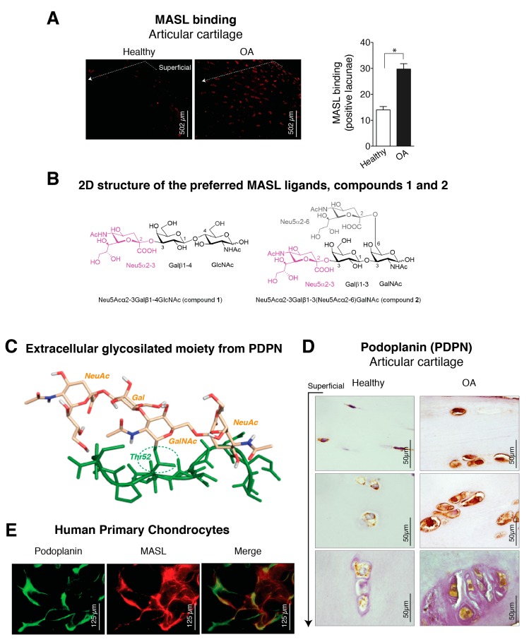
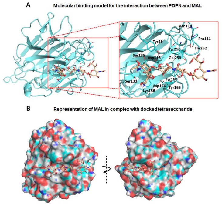
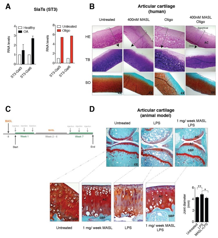
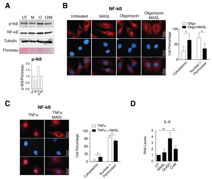
Osteoarthritis (OA) is the most common degenerative joint disease characterized by articular cartilage degradation and joint degeneration. The articular cartilage is mainly formed by chondrocytes and a collagen-proteoglycan extracellular matrix that contains high levels of glycosylated proteins. It was reported that the shift from glycoproteins containing α-2,6-linked sialic acids to those that contain α-2,3 was associated with the onset of common types of arthritis. However, the pathophysiology of α-2,3-sialylation in cartilage has not been yet elucidated. We show that cartilage from osteoarthritic patients expresses high levels of the α-2,3-sialylated transmembrane mucin receptor, known as podoplanin (PDPN). Additionally, the Maackia amurensis seed lectin (MASL), that can be utilized to target PDPN, attenuates the inflammatory response mediated by NF-kB activation in primary chondrocytes and protects human cartilage breakdown ex vivo and in an animal model of arthritis. These findings reveal that specific lectins targeting α-2,3-sialylated receptors on chondrocytes might effectively inhibit cartilage breakdown. We also present a computational 3D molecular model for this interaction. These findings provide mechanistic information on how a specific lectin could be used as a novel therapy to treat degenerative joint diseases such as osteoarthritis.

Keywords: MASL; arthritis; articular chondrocyte; cartilage; glycoproteins; molecular modelling; osteoarthritis; podoplanin; sialylation.


Conflict of interest statement

The authors declare no competing financial interests.

Figures

Figure A1
Figure A1
MASL treatment does not affect chondrocyte cell viability, adhesion, growth, or migration. (a) Cells from osteoarthritis patients were cultured for 1 or 17 h with 0, 400, and 720 nM MASL with or without 5 µg/mL of oligomycin as indicated. Cytotoxicity was evaluated with the colorimetric MTT assay, and the data are presented as the mean ± SD (n = 2). (b) Confluent monolayers of primary human chondrocytes, TC28a2 cells (a human chondrocyte cell line), and primary human synoviocytes were treated for 1 h with varying concentrations of MASL as indicated. Cell adhesion was evaluated with fibrinogen-coated plates, with BSA-coated plates used as controls. The data are shown as the mean ± SD (n = 2). (c) Chondrocytes were grown to confluence in wells containing an insert that forms a 0.9 mm gap in the monolayer. After the insert was removed, the cells were treated for 24 h (TC28a2 cells) with 400 or 720 nM MASL or 10 days (primary chondrocytes) and imaged under an inverted light microscope. No differences in cell adhesion or growth were observed between untreated and treated samples (n = 4).
Figure A2
Figure A2
Molecular binding model for the interaction between the PDPN tetrasaccharide 2 and MAH. The MAH homology model (Ref. Q7M1M0, UniProt) was generated with the Phyre2 web portal server [83] sing MAL (Ref. P0DKL3, UniProt) as a template, with a query cover of 95% and a percentage of identity of more than 80% (Table A1). Docked tetrasaccharide 2 (Neu5Acα(2–3)Galβ-(1–3)[Neu5Acα(2–6)]GalNAc, shown in beige) in complex with MAH (magenta). (A) Selected residues participating in the main interactions are displayed in sticks and labels are shown and (B) Surface representation of MAH in complex with docked tetrasaccharide 2. The electrostatic potential is coloured on the surface of the protein from red (negative) to blue (positive).
Figure A3
Figure A3
MASL protects articular cartilage structure in vivo in pathological conditions: animal model. Images show representative samples of haematoxylin–eosin (HE), toluidine blue, and Safranin-O Fast Green staining (see Figure 3D).
Figure 1
Figure 1
Expression of α-2,3-sialylated glycoproteins, represented by podoplanin (PDPN), is induced in arthritic chondrocytes. (A) Maackia amurensis seed lectin (MASL) conjugated to HiLyte Fluor TR (red) was used to detect α-2,3-sialylated glycoproteins in cartilage sections from healthy donors (n = 4) and patients with grade II osteoarthritis (OA) (n = 4). The graph represents the number of positive lacunae (mean ± SEM), n = 4, * p < 0.05, Mann–Whitney test. Healthy donors are represented by white bar and OA patients in black; (B) 2D structure of the preferred Maackia amurensis ligands. Left: sialyllactosamine (compound 1), preferred by leucoagglutinin (MAL). Right: tetrasaccharide (compound 2), preferred by haemagglutinin (MAH). Both sugars contain α-2,3 or/and α-2,6 sialyl (Neu5Ac) linkages (in magenta Neu5Acα2–3 and in grey Neu5Acα2–6); (C) 3D structure of the extracellular glycosylated moiety of PDPN adapted from PDBID 3WSR. The peptidic portion is shown in green. Thr52 is O-glycosylated with Neu5Acα(2–3)Galβ-(1–3)[Neu5Acα(2–6)]GalNAc, which is shown in beige; (D) PDPN protein levels were detected by IHC analysis of cartilage from healthy donors (n = 4) and grade II patients with OA (n = 9). See Figure A1. Arrow indicates cartilage sections extending from the superficial, adjacent to the synovial fluid, into the intermediate and deep zones; (E) Primary chondrocytes from healthy donors and grade II OA patients were incubated with HiLyte Fluor TR-labelled MASL to detect α-2,3-sialylated glycoproteins (red) and with monoclonal antibody to detect PDPN by immunofluorescence (green). Colocalization of MASL and PDPN was evident in the merged images (yellow).
Figure 2
Figure 2
Molecular binding model for the interaction between the PDPN tetrasaccharide 2 and Maackia amurensis seed lectin (MAL): Docked tetrasaccharide 2 (Neu5Acα(2–3)Galβ-(1–3)[Neu5Acα(2–6)]GalNAc, in beige) in complex with MAL (cyan). (A) Selected residues participating in the main interactions are displayed in sticks and labels and (B) surface representation of MAL in complex with docked tetrasaccharide 2. The electrostatic potential is coloured on the surface of the protein from red (negative) to blue (positive).
Figure 3
Figure 3
MASL protects cartilage matrix from LPS-induced degeneration. (A) mRNA levels of the sialyltransferases ST3-Gal3 and ST3-Gal6 in primary chondrocytes obtained from healthy donors and OA patients. The data are presented as the mean ± SEM, n = 3 (p = 0.6579 and p = 0.0765 for healthy donors and OA patients, respectively, Mann–Whitney test). mRNA levels of ST3-Gal3 and ST3-Gal6 in cartilage explants exposed to 5 µg/mL of oligomycin for 7 days. The data are presented as the mean ± SD (n = 2). (B) Histological sections of 4 mm circular biopsy punches that were cultured for 7 days with or without MASL and oligomycin. Human cartilage punches (4 mm) from osteoarthritis patients treated with MASL (400 nM) and 5 µg/mL of oligomycin for 7 days. Sections were stained for haematoxylin–eosin (HE), toluidine blue (TB), and Safranine-O Fast Green (SO). Cartilage damage resulting from extracellular matrix (ECM) degradation is shown. As it has been previously observed, MASL prevented the loss of ECM components, tissue degradation, and increases in lacuna spaces [43]. Arrows indicate cartilage sections extending from superficial, adjacent to synovial fluid, into the intermediate and deep zones. (C) Schematic representation of the in vivo model workflow. (D) Staining of knee joints with Safranin-O Fast Green exhibits histological changes in the subchondral bone plate (SBP) and articular cartilage (AC) of the femorotibial joints of mice in the LPS-induced arthritis model. LPS-induced arthritic mice, which were treated with 1 mg of MASL per week for 7 weeks, did not show significant changes in subchondral bone and cartilage structure in comparison with controls (Table 1). Knee joint diameters (in mm) measured 24 h after LPS injection with or without MASL treatment (48 h after oral administration of MASL) are shown in the graph. The data are presented as the mean ± SEM (n = 6), * p < 0.05, ** p < 0.01; Mann–Whitney test.
Figure 4
Figure 4
MASL attenuates nuclear factor kappa B p65 activation. (A) Western blot analysis of phospho-IkB-α and NF-kB in primary chondrocytes in monolayer culture, untreated (U) and treated with 400 nM MASL (M and O/M) or 5 µg/mL of oligomycin (O) for 1 h. α-tubulin and ponceau staining were used as loading controls. p-Ikß quantification is shown below. Representative experiment of n = 3. (B) Detection of NF-kB (p65) in red by immunofluorescence. Nuclei were stained with DAPI. Primary chondrocytes were treated for 1 h with MASL (400 nM) and 5 µg/mL oligomycin. Scale bar, 50 µm. Quantification is shown on the right. Values indicate percentage of cells. The data are presented as the mean ± SEM (n = 3), * p < 0.05; Mann–Whitney test. (C) Detection of NF-kB (p65) by immunofluorescence in primary chondrocytes treated with 10 ng/mL TNF-α and 400 nM MASL for 1 h. Scale bar, 50 µm. Quantification is shown on the right. Values indicate percentage of cells. The data are presented as the mean ± SEM (n = 3), * p < 0.05; Mann–Whitney test; (D) mRNA levels of IL-6 in primary chondrocytes exposed to MASL (400 nM) and/or 5 µg/mL of oligomycin for 1 h. The data are presented as the mean ± SEM (n = 3). * p < 0.05, **p < 0.01; Mann–Whitney test.

References

    1. Loeser R.F., Goldring S.R., Scanzello C.R., Goldring M.B. Osteoarthritis: A disease of the joint as an organ. Arthritis Rheum. 2012;64:1697–1707. doi: 10.1002/art.34453. - DOI - PMC - PubMed
    1. Moremen K.W., Tiemeyer M., Nairn A.V. Vertebrate protein glycosylation: Diversity, synthesis and function. Nat. Rev. Mol. Cell Biol. 2012;13:448–462. doi: 10.1038/nrm3383. - DOI - PMC - PubMed
    1. Turner G.A. N-glycosylation of serum proteins in disease and its investigation using lectins. Clin. Chim. Acta. 1992;208:149–171. doi: 10.1016/0009-8981(92)90073-Y. - DOI - PubMed
    1. Toegel S., Pabst M., Wu S.Q., Grass J., Goldring M.B., Chiari C., Kolb A., Altmann F., Viernstein H., Unger F.M. Phenotype-related differential alpha-2,6- or alpha-2,3-sialylation of glycoprotein N-glycans in human chondrocytes. Osteoarthr. Cartil. 2010;18:240–248. doi: 10.1016/j.joca.2009.09.004. - DOI - PMC - PubMed
    1. Schultz M.J., Swindall A.F., Bellis S.L. Regulation of the metastatic cell phenotype by sialylated glycans. Cancer Metastasis Rev. 2012;31:501–518. doi: 10.1007/s10555-012-9359-7. - DOI - PMC - PubMed

Publication types

MeSH terms