. 2020 Apr 15;21(8):2734.
doi: 10.3390/ijms21082734.

Acute and Chronic Dopaminergic Depletion Differently Affect Motor Thalamic Function

Affiliations

Acute and Chronic Dopaminergic Depletion Differently Affect Motor Thalamic Function

Giuseppe Di Giovanni et al. Int J Mol Sci. .

Abstract

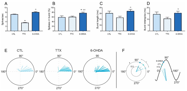
The motor thalamus (MTh) plays a crucial role in the basal ganglia (BG)-cortical loop in motor information codification. Despite this, there is limited evidence of MTh functionality in normal and Parkinsonian conditions. To shed light on the functional properties of the MTh, we examined the effects of acute and chronic dopamine (DA) depletion on the neuronal firing of MTh neurons, cortical/MTh interplay and MTh extracellular concentrations of glutamate (GLU) and gamma-aminobutyric acid (GABA) in two states of DA depletion: acute depletion induced by the tetrodotoxin (TTX) and chronic denervation obtained by 6-hydroxydopamine (6-OHDA), both infused into the medial forebrain bundle (MFB) in anesthetized rats. The acute TTX DA depletion caused a clear-cut reduction in MTh neuronal activity without changes in burst content, whereas the chronic 6-OHDA depletion did not modify the firing rate but increased the burst firing. The phase correlation analysis underscored that the 6-OHDA chronic DA depletion affected the MTh-cortical activity coupling compared to the acute TTX-induced DA depletion state. The TTX acute DA depletion caused a clear-cut increase of the MTh GABA concentration and no change of GLU levels. On the other hand, the 6-OHDA-induced chronic DA depletion led to a significant reduction of local GABA and an increase of GLU levels in the MTh. These data show that MTh is affected by DA depletion and support the hypothesis that a rebalancing of MTh in the chronic condition counterbalances the profound alteration arising after acute DA depletion state.

Keywords: L-DOPA; deep brain stimulation; electrophysiology; immunohistochemistry; microdialysis.


Conflict of interest statement

The authors declare no conflict of interest.

Figures

Figure 1
Figure 1
(A) Representative electrophysiological recordings of the three groups of animals. From the Table 6. hydroxydopamine (6-OHDA)-lesioned rats, respectively. (C) Histological reconstruction of the MTh recording site in tetrodotoxin (TTX)-treated rats (in blue). The black circles represent the recorded neurons in MTh during the TTX-mediated blockade of MFB.
Figure 2
Figure 2
(AD) Firing rate and pattern of the MTh in CTL, during tetrodotoxin (TTX) infusion and in 6-hydroxydopamine (6-OHDA)-lesioned rats. * p < 0.05. (E,F). Phase correlation analysis. (E) MTh circular plots of the phase-locked firing of the MTh neurons in control, acute and chronic DA depletion states. The vectors of the preferred firing of individual neurons are shown as lines radiating from the center. The last smaller circular plots show the mean vector for the preferred phases of neurons in each group. (F) The histograms on the right show the vector lengths and phase angle comparisons between MTh (blue) in control (dark color), acute (dashed color) and chronic (light color) DA depletion states. On the left, the representation of hypothetical cortical slow oscillation comparing the phase angle and vector length between MTh (blue) control (dark color), TTX (dashed color) and 6-OHDA (light color) rats. * p < 0.05, Kruskal–Wallis test. MTh: motor thalamus; CTL: control; TTX: tetrodotoxin; 6-OHDA: 6-hydroxydopamine. The 0° represents the mid-ascending phase of cortical slow-wave activity (SWA), the 90° represents the cortical SWA peak and the 180° represents the mid-descending phase of cortical SWA.
Figure 3
Figure 3
Effect of acute (TTX) and chronic (6-OHDA) dopamine (DA)-depletion on the extracellular levels of endogenous GABA and glutamate (GLU) in the motor thalamus (MTh). TTX infused in the medial forebrain bundle induced an increase in the endogenous GABA extracellular levels (A) while did not change the GLU levels in the MTh. Data in (A) and (B) are expressed as mean ± SEM normalized to the baseline levels (pre TTX infusion) from n = 14 experiments per group; * p < 0.05. One-and two-way ANOVA for repeated measures, followed by Dunnet’s multiple comparison test. 6-OHDA-lesion infused in the medial forebrain bundle produced, after 2 weeks, a decrease in GABA (C) and an increase in GLU extracellular levels in the MTh (D). Data in (C) and (D) are expressed as means ± SEMs normalized to sham-lesioned (n = 9) neurotransmitter levels from n = 9 experiments per group; ** p < 0.01, unpaired student t-test.

References

    1. Bosch-Bouju C., Hyland B.I., Parr-Brownlie L.C. Motor thalamus integration of cortical, cerebellar and basal ganglia information: Implications for normal and parkinsonian conditions. Front. Comput. Neurosci. 2013;7:163. doi: 10.3389/fncom.2013.00163. - DOI - PMC - PubMed
    1. Nakamura K.C., Sharott A., Magill P.J. Temporal coupling with cortex distinguishes spontaneous neuronal activities in identified basal ganglia-recipient and cerebellar-recipient zones of the motor thalamus. Cereb. Cortex. 2014;24:81–97. doi: 10.1093/cercor/bhs287. - DOI - PMC - PubMed
    1. Wichmann T., Kliem M.A. Neuronal activity in the primate substantia nigra pars reticulata during the performance of simple and memory-guided elbow movements. J. Neurophysiol. 2004;91:815–827. doi: 10.1152/jn.01180.2002. - DOI - PubMed
    1. Avila I., Parr-Brownlie L.C., Brazhnik E., Castaneda E., Bergstrom D.A., Walters J.R. Beta frequency synchronization in basal ganglia output during rest and walk in a hemiparkinsonian rat. Exp. Neurol. 2010;221:307–319. doi: 10.1016/j.expneurol.2009.11.016. - DOI - PMC - PubMed
    1. Pare D., Steriade M., Deschenes M., Oakson G. Physiological characteristics of anterior thalamic nuclei, a group devoid of inputs from reticular thalamic nucleus. J. Neurophysiol. 1987;57:1669–1685. doi: 10.1152/jn.1987.57.6.1669. - DOI - PubMed

MeSH terms

LinkOut - more resources