Review
. 2020 Apr 15;9(4):313.
doi: 10.3390/antiox9040313.

Oxidative Stress, a Crossroad Between Rare Diseases and Neurodegeneration

Affiliations
Review

Oxidative Stress, a Crossroad Between Rare Diseases and Neurodegeneration

Carmen Espinós et al. Antioxidants (Basel). .

Abstract

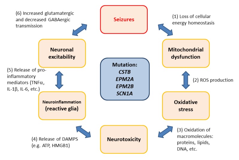
: Oxidative stress is an imbalance between production and accumulation of oxygen reactive species and/or reactive nitrogen species in cells and tissues, and the capacity of detoxifying these products, using enzymatic and non-enzymatic components, such as glutathione. Oxidative stress plays roles in several pathological processes in the nervous system, such as neurotoxicity, neuroinflammation, ischemic stroke, and neurodegeneration. The concepts of oxidative stress and rare diseases were formulated in the eighties, and since then, the link between them has not stopped growing. The present review aims to expand knowledge in the pathological processes associated with oxidative stress underlying some groups of rare diseases: Friedreich's ataxia, diseases with neurodegeneration with brain iron accumulation, Charcot-Marie-Tooth as an example of rare neuromuscular disorders, inherited retinal dystrophies, progressive myoclonus epilepsies, and pediatric drug-resistant epilepsies. Despite the discrimination between cause and effect may not be easy on many occasions, all these conditions are Mendelian rare diseases that share oxidative stress as a common factor, and this may represent a potential target for therapies.

Keywords: Charcot-Marie-Tooth disease (CMT); Dravet syndrome; Friedreich’s ataxia; Lafora disease (LD); Unverricht–Lundborg disease (ULD); inherited retinal dystrophy (IRD); neurodegenerative disorders with brain iron accumulation (NBIA); progressive myoclonus epilepsy (PME).


Conflict of interest statement

The authors declare no conflict of interest.

Figures

Figure 1
Figure 1
Main mechanisms in ferroptosis. (A) General process of ferroptosis. Balanced levels of reactive oxygen species (ROS) generation and antioxidant activity maintains an inactive ferroptosis cell death pathway; however, some disturbance in the players of this equilibrium could produce a ferroptosis activation. Two important components of ferroptosis are the process of transferrin (TF)-iron import and ferroportin (FPN1) export. Changes in iron uptake or iron export produce a suppression or activation of ferroptosis since the free iron pool is fundamental [31,32]. Ferroptosis may be enhanced through the process of ferritinophagy, by which ferritin is targeted by nuclear receptor coactivator 4 (NCOA4) and delivered to the lysosome for degradation in autophagosome [33,34,35]. Consequently, a large quantity of iron can be rapidly released. Currently, it has been suggested that ferroptosis initiation might be triggered by an increase in free iron levels itself [36]. Iron increase/or accumulation induces the Fenton reaction which increases the production of ROS. Furthermore, the action of iron-dependent oxidases [37], specifically lipoxygenase activity of 15-LOX (ALOX15) that oxidizes polyunsaturated fatty acids (PUFAs) phospholipids (PUFA-PLs) containing arachidonate or adrenate moieties, triggers the ferroptosis pathway [38,39]. In addition, increased activity of acyl-CoA synthetase long-chain family member 4 (ACSL4), an enzyme that preferentially activates arachidonic (AA) and adrenoic acids (AdA), has been reported to increase ferroptosis [38,40]. Inhibition of glutathione peroxidase enzyme 4 (GPX4) [2,3] or glutathione (GSH) unavailability [4,5] produces lipid hydroperoxide accumulation that triggers ferroptosis. (B) Suggested mechanisms of ferroptosis in Friedreich’s ataxia (FRDA). It has been observed increased levels of lipoperoxides and ROS in FRDA neurons. Furthermore, in these neurons, it has also been reported lower reduced GSH concentration and GSH homeostasis disturbance, respectively [41,42,43]. Controversial studies are reporting either normal or increased iron levels in the nervous system of FRDA patients (reviewed in [44]). Recently a study in FRDA patients, using magnetic resonance imaging, showed increased iron concentration in the extrapyramidal motor system compared with controls [45]. However, there is no information about distribution, location and redox state of the iron in cells from FRDA (indicated with “?”). Increased autophagic processes are related to FRDA in different models of disease [46,47], but there is no information about ferritinophagic activity. Further studies should be performed in order to unravel if this mechanism increases the iron content in FRDA cells. All these ferroptotic characteristics observed in FRDA may indicate that this kind of cell death may have a role in the physiopathology of this disease. Transferrin receptor (TFR) Lysophosphatidylcholine Acyltransferase 3 (LPCAT3); Glutamate Cysteine Ligase (GCL); Glutathione synthetase (GSS); Glutathione reductase (GR). ↑ indicates increased levels and ↓ indicates decreased levels.
Figure 2
Figure 2
Pathways underlying NBIA disorders. PANK2 and COASY (in green) are involved in the coenzyme A (CoA) synthesis. PLA2G6, C19orf12, FA2H, SCP2, and CRAT (in purple) are related to lipid metabolism and membrane remodeling. CP and FTL1 (in orange) are implicated in iron metabolism; WDR45 and ATP13A2 (in maroon) play a role in autophagy; and finally, REPS1 and AP4M1 (in blue) are associated with vesicle trafficking. The function of DCAF17 and GTPBP2 (in fuchsia) remains elusive.
Figure 3
Figure 3
Charcot-Marie-Tooth disease (CMT) pathways related to oxidative stress. CMT is clinically divided into three main types, the demyelinating form, characterized by a decrease in the nervous conduction velocity; axonal CMT, where there is a loss in the number of axons and an intermediate clinical form with some decrease in the velocity of conduction and a decrease in the number of axons. At least 80 different genes are affected in the disease. Some of them produce oxidative stress or mitochondrial impairment. MFN2 and GDAP1 are the best known. HSPB8 has been related to the Nrf2 pathway. ER: Endoplasmic reticulum, GDAP1: ganglioside induced differentiation associated protein 1, GSH: glutathione, HSPB8: Small Heat shock protein 8, MFN2: mitofusin 2, Nrf2: Nuclear factor erythroid 2-related factor 2, ROS: reactive oxygen species.
Figure 4
Figure 4
Therapeutic approaches for retinitis pigmentosa (RP), the most prevalent inherited retinal dystrophy. RP is characterized by progressive rod photoreceptor degeneration (mutated genes) in the initial stage and eventual cone photoreceptor degeneration in later stages. It is highly probable that cone degeneration is influenced by the release of inflammatory molecules, oxidant radicals, pro-apoptotic factors, etc. from rods and other cells, independently of the gene defect. Several therapeutic approaches have been developed to preserve retinal function or restore vision depending on the stage of the disease. They can be led to repair mutated photoreceptors (gene therapy), to replace damaged photoreceptors (cell therapy), or to protect photoreceptors and other retinal cells to the “toxic environment” (neuroprotective compounds including antioxidants). At later stages of RP, therapeutic approaches can be led to other retinal cells to restore visual activity with the remaining retinal circuits (optogenetics, retinal prostheses).
Figure 5
Figure 5
The vicious circle of oxidative stress and neuroinflammation in rare epilepsies with a genetic origin. (1) There is an inverse relationship between mitochondria functionality and the appearance of seizures. Alterations in cellular energy homeostasis result in neuronal pathology. (2) Mitochondrial dysfunction leads to oxidative stress due to the accumulation of reactive oxygen species (ROS). (3) Oxidized macromolecules (e.g., proteins, lipids, DNA) affect neuronal viability, leading to neurotoxicity. (4) Neuronal release of danger associated molecular patterns (DAMPs; e.g., HMGB1, ATP) triggers a neuroinflammatory response, leading to the activation of glia (microglia and astrocytes). (5) The production of pro-inflammatory mediators (TNFα, IL-1β, IL-6, etc.) affects neuronal excitability. (6) Increased glutamatergic transmission and decreased GABAergic inhibition leads to seizures.

References

    1. Sies H. Oxidative stress: A concept in redox biology and medicine. Redox Biol. 2015;4:180–183. doi: 10.1016/j.redox.2015.01.002. - DOI - PMC - PubMed
    1. Gaschler M.M., Stockwell B.R. Lipid peroxidation in cell death. Biochem. Biophys. Res. Commun. 2017;482:419–425. doi: 10.1016/j.bbrc.2016.10.086. - DOI - PMC - PubMed
    1. Yang W.S., Kim K.J., Gaschler M.M., Patel M., Shchepinov M.S., Stockwell B.R. Peroxidation of polyunsaturated fatty acids by lipoxygenases drives ferroptosis. Proc. Natl. Acad. Sci. USA. 2016;113:E4966–E4975. doi: 10.1073/pnas.1603244113. - DOI - PMC - PubMed
    1. Cao J.Y., Poddar A., Magtanong L., Lumb J.H., Mileur T.R., Reid M.A., Dovey C.M., Wang J., Locasale J.W., Stone E., et al. A Genome-wide Haploid Genetic Screen Identifies Regulators of Glutathione Abundance and Ferroptosis Sensitivity. Cell Rep. 2019;26:1544–1556. doi: 10.1016/j.celrep.2019.01.043. - DOI - PMC - PubMed
    1. Dixon S.J., Lemberg K.M., Lamprecht M.R., Skouta R., Zaitsev E.M., Gleason C.E., Patel D.N., Bauer A.J., Cantley A.M., Yang W.S., et al. Ferroptosis: An iron-dependent form of nonapoptotic cell death. Cell. 2012;149:1060–1072. doi: 10.1016/j.cell.2012.03.042. - DOI - PMC - PubMed