. 2020 Jul;98(1):219-227.
doi: 10.1016/j.kint.2020.04.003. Epub 2020 Apr 9.

Renal histopathological analysis of 26 postmortem findings of patients with COVID-19 in China

Affiliations

Renal histopathological analysis of 26 postmortem findings of patients with COVID-19 in China

Hua Su et al. Kidney Int. 2020 Jul.

Abstract

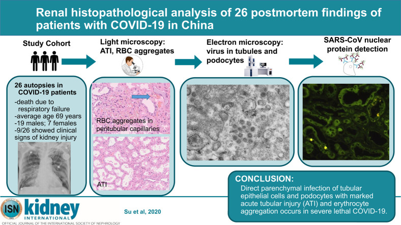
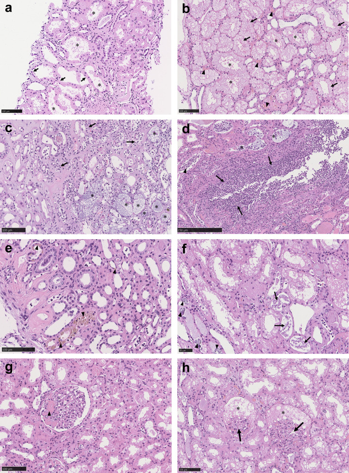
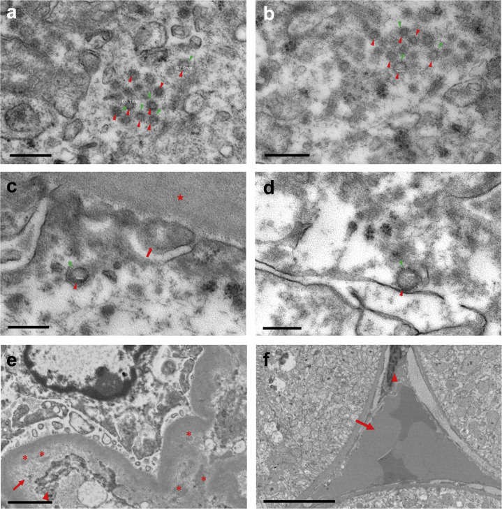
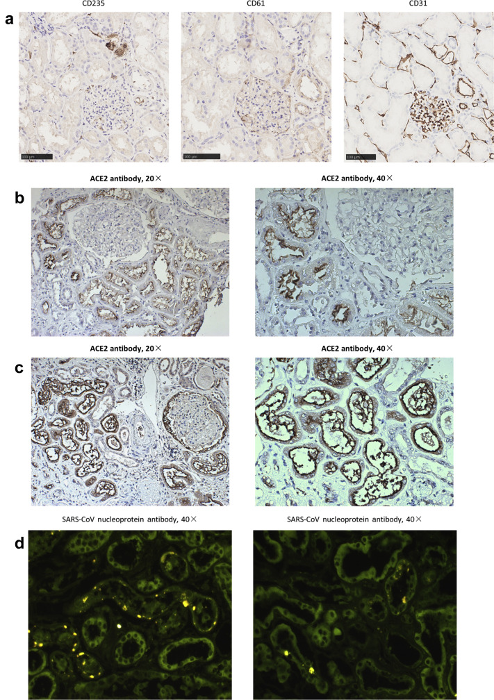
Although the respiratory and immune systems are the major targets of Coronavirus Disease 2019 (COVID-19), acute kidney injury and proteinuria have also been observed. Currently, detailed pathologic examination of kidney damage in critically ill patients with COVID-19 has been lacking. To help define this we analyzed kidney abnormalities in 26 autopsies of patients with COVID-19 by light microscopy, ultrastructural observation and immunostaining. Patients were on average 69 years (19 male and 7 female) with respiratory failure associated with multiple organ dysfunction syndrome as the cause of death. Nine of the 26 showed clinical signs of kidney injury that included increased serum creatinine and/or new-onset proteinuria. By light microscopy, diffuse proximal tubule injury with the loss of brush border, non-isometric vacuolar degeneration, and even frank necrosis was observed. Occasional hemosiderin granules and pigmented casts were identified. There were prominent erythrocyte aggregates obstructing the lumen of capillaries without platelet or fibrinoid material. Evidence of vasculitis, interstitial inflammation or hemorrhage was absent. Electron microscopic examination showed clusters of coronavirus-like particles with distinctive spikes in the tubular epithelium and podocytes. Furthermore, the receptor of SARS-CoV-2, ACE2 was found to be upregulated in patients with COVID-19, and immunostaining with SARS-CoV nucleoprotein antibody was positive in tubules. In addition to the direct virulence of SARS-CoV-2, factors contributing to acute kidney injury included systemic hypoxia, abnormal coagulation, and possible drug or hyperventilation-relevant rhabdomyolysis. Thus, our studies provide direct evidence of the invasion of SARSCoV-2 into kidney tissue. These findings will greatly add to the current understanding of SARS-CoV-2 infection.

Keywords: COVID-19; SARS-CoV-2; acute kidney injury; proteinuria; renal pathology.


Figures

None
Graphical abstract
Figure 1
Figure 1
Spectrum of pathologic abnormalities of kidneys from postmortems of patients with coronavirus disease 2019. (a,b) Proximal tubules showed (a) loss of brush border and (b) vacuolar degeneration (arrows), with debris composed of necrotic epithelium in tubular lumens (asterisks). Erythrocyte aggregates obstructing peritubular capillaries were frequently present (arrowheads). (c,d) Some cases showed infiltration of inflammatory cells in (c) tubules and (d) in 1 case, in an arcuate artery (arrows), with multiple foci of bacteria (asterisks) and white blood cell casts (arrowhead). (e,f) Occasional (e) hemosiderin granules and (f) deposits of calcium (arrowheads) were present in tubules with occasional pigmented casts (arrows). (g,h) Segmental fibrin thrombi were present in glomeruli (arrowhead), with ischemic glomerular contraction (arrows) with the accumulation of leaked plasma in Bowman’s space (asterisks). Hematoxylin and eosin. Bars = (f) 50 μm, (a–c,e,g,h) 100 μm, and (d) 250 μm. To optimize viewing of this image, please see the online version of this article at www.kidney-international.org.
Figure 2
Figure 2
Ultrastructural features of kidneys from postmortems of patients with coronavirus disease 2019. (a–d) Coronavirus-like particles (red arrowheads) with distinctive spikes (green arrowheads) were present in the cytoplasm of (a) the proximal and (b) distal tubular epithelium. (c,d) Coronavirus-like particles (red arrowheads) with distinctive spikes (green arrowheads) were present in podocytes; foot processes of podocytes (arrow); glomerular basement membrane (star). (e) A single case of IgA nephropathy was diagnosed by immunofluorescent staining with a few paramesangial and subendothelial electron-dense deposits (asterisks) with marked subendothelial lucent expansion (arrow) and mesangial interposition (arrowhead). (f) Peritubular capillary with stasis of red blood cells (arrow) and activation or injury of endothelial cells (arrowhead). Transmission electron microscopy. Bars = (a–d) 200 nm, (e) 1 μm, and (f) 5 μm. To optimize viewing of this image, please see the online version of this article at www.kidney-international.org.
Figure 3
Figure 3
Immunostaining of paraffin-embedded kidney tissue from patients with coronavirus disease 2019 (COVID-19). (a) Serial sections stained for CD235, CD61, and CD31 showing stasis of red blood cells without platelets in peritubular capillaries. Bars = 100 μm. (b,c) Angiotensin-converting enzyme II (ACE2) stained mainly proximal tubules in (b) noncoronavirus disease 2019 case, with (c) strong proximal tubular staining and parietal epithelial cell staining with occasional weak podocyte staining in some COVID-19 cases. (d) Indirect immunofluorescent staining with anti–severe acute respirator syndrome coronavirus (SARS-CoV) nucleoprotein antibody. To optimize viewing of this image, please see the online version of this article at www.kidney-international.org.

Comment in

  • COVID-19-associated nephritis: early warning for disease severity and complications?
    Gross O, Moerer O, Weber M, Huber TB, Scheithauer S. Gross O, et al. Lancet. 2020 May 16;395(10236):e87-e88. doi: 10.1016/S0140-6736(20)31041-2. Epub 2020 May 6. Lancet. 2020. PMID: 32423587 Free PMC article. No abstract available.
  • The authors reply.
    Su H, Gao D, Yang HC, Fogo AB, Nie X, Zhang C. Su H, et al. Kidney Int. 2020 Jul;98(1):232-233. doi: 10.1016/j.kint.2020.05.007. Epub 2020 May 16. Kidney Int. 2020. PMID: 32425266 Free PMC article. No abstract available.
  • Visualization of putative coronavirus in kidney.
    Miller SE, Brealey JK. Miller SE, et al. Kidney Int. 2020 Jul;98(1):231-232. doi: 10.1016/j.kint.2020.05.004. Epub 2020 May 8. Kidney Int. 2020. PMID: 32437764 Free PMC article. No abstract available.
  • Am I a coronavirus?
    Smith KD, Akilesh S, Alpers CE, Nicosia RF. Smith KD, et al. Kidney Int. 2020 Aug;98(2):506-507. doi: 10.1016/j.kint.2020.05.021. Epub 2020 Jun 4. Kidney Int. 2020. PMID: 32505464 Free PMC article. No abstract available.

References

    1. Lu R., Zhao X., Li J. Genomic characterisation and epidemiology of 2019 novel coronavirus: implications for virus origins and receptor binding. Lancet. 2020;395:565–574. - PMC - PubMed
    1. Guan W.J., Ni Z.Y., Hu Y. Clinical characteristics of coronavirus disease 2019 in China. N Engl J Med. 2020;382:1708–1720. - PMC - PubMed
    1. Alsaad K.O., Hajeer A.H., Al Balwi M. Histopathology of Middle East respiratory syndrome coronavirus (MERS-CoV) infection—clinicopathological and ultrastructural study. Histopathology. 2018;72:516–524. - PMC - PubMed
    1. Xu Z., Shi L., Wang Y. Pathological findings of COVID-19 associated with acute respiratory distress syndrome. Lancet Respir Med. 2020;8:420–422. - PMC - PubMed
    1. Tietäväinen J., Mantula P., Outinen T. Glucosuria predicts the severity of Puumala hantavirus infection. Kidney Int Rep. 2019;4:1296–1303. - PMC - PubMed

Publication types