. 2020 May;41(5):923-928.
doi: 10.3174/ajnr.A6532. Epub 2020 Apr 23.

Cerebellar Watershed Injury in Children

Affiliations

Cerebellar Watershed Injury in Children

J N Wright et al. AJNR Am J Neuroradiol. 2020 May.

Abstract

Background and purpose: Focal signal abnormalities at the depth of the cerebellar fissures in children have recently been reported to represent a novel pattern of bottom-of-fissure dysplasia. We describe a series of patients with a similar distribution and appearance of cerebellar signal abnormality attributable to watershed injury.

Materials and methods: Twenty-three children with MR imaging findings of focal T2 prolongation in the cerebellar gray matter and immediate subjacent white matter at the depth of the fissures were included. MR imaging examinations were qualitatively analyzed for the characteristics and distribution of signal abnormality within posterior fossa structures, the presence and distribution of volume loss, the presence of abnormal contrast enhancement, and the presence and pattern of supratentorial injury.

Results: T2 prolongation was observed at the depths of the cerebellar fissures bilaterally in all 23 patients, centered at the expected location of the deep cerebellar vascular borderzone. Diffusion restriction was associated with MR imaging performed during acute injury in 13/16 patients. Five of 23 patients had prior imaging, all demonstrating a normal cerebellum. The etiology of injury was hypoxic-ischemic injury in 17/23 patients, posterior reversible encephalopathy syndrome in 3/23 patients, and indeterminate in 3/23 patients. Twenty of 23 patients demonstrated an associated classic parasagittal watershed pattern of supratentorial cortical injury. Injury in the chronic phase was associated with relatively preserved gray matter volume in 8/15 patients, closely matching the published appearance of bottom-of-fissure dysplasia.

Conclusions: In a series of patients with findings similar in appearance to the recently described bottom-of-fissure dysplasia, we have demonstrated a stereotyped pattern of injury attributable to cerebellar watershed injury.


Figures

Fig 1.
Fig 1.
Axial T2WI from patient 2 (A), parasagittal T2WI from patient 8 (B), and a coronal T2WI from patient 6 (C) demonstrate the characteristic appearance and location of cerebellar watershed injury. There is T2 prolongation greatest in the gray matter at the depths of the cerebellar fissures (white arrows) within the deep cerebellar watershed territory.
Fig 2.
Fig 2.
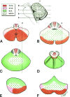
Schematic drawing of the vascular territories of the cerebellum. Note the central distribution of the cerebellar watershed areas (WSCA) at the junction of PICA, AICA, and SCA. The distribution of MR imaging signal abnormality in our case series corresponds to cerebellar watershed regions, best demonstrated by comparing the axial (B), parasagittal (E), and coronal (F) schematic images with multiplanar MR images in Fig 1. Modified from Savoiardo et al.
Fig 3.
Fig 3.
Coronal T2WI from patient 4 (A, C, and E) and patient 11 (B, D, and F) demonstrates the appearance of newly developed acute (C and D) and subsequent chronic (E and F) phase injury in previously normal cerebella (A and B). White arrows highlight multipe sites demonstrating progressive development of typical watershed injury in previously normal gray matter.
Fig 4.
Fig 4.
Coronal T2WI (A), coronal DWI (B), and coronal ADC map (C) from patient 18 demonstrate acute cerebellar and supratentorial injury with T2 prolongation and restricted diffusion in the setting of HII. Coronal T2WI (D), coronal DWI (E), and coronal ADC map (F) from patient 9 demonstrate acute cerebellar (white arrowheads) and supratentorial (white arrows) injury without diffusion restriction in the setting of PRES.
Fig 5.
Fig 5.
Coronal T1WI from patient 22 in the subacute (A) and chronic (B) phases demonstrates focal T1 hyperintensity in the bilateral cerebellar watershed (white arrows in A), consistent with laminar necrosis and resolved within 1 month on follow-up (white arrows in B).
Fig 6.
Fig 6.
Coronal T2WI from patients during the chronic phase. Patients 1 (A) and 2 (B) demonstrate T2 prolongation with relatively marked gray matter volume loss at the sites of prior injury (white arrows). Patients 7 (C) and 10 (D) demonstrate a thick lamina of gray matter T2 prolongation centered at the depths of the cerebellar fissures in an arterial watershed distribution. See also Figs 1C, 3E, and for additional examples.
Fig 7.
Fig 7.
Findings of both acute and remote supratentorial watershed injury in patients 1–6 (AF, respectively). Acute watershed infarcts with signal abnormality on axial (A and B) and coronal (C) DWI in a characteristic parasagittal distribution. Subacute or remote watershed infarcts with hyperintensity and parenchymal volume loss on coronal (D and F) and axial (E) T2.

Comment in

  • Reply.
    Wright JN, Shaw DWW, Ishak G, Perez FA, Doherty D. Wright JN, et al. AJNR Am J Neuroradiol. 2020 Aug;41(8):E61. doi: 10.3174/ajnr.A6673. Epub 2020 Jun 25. AJNR Am J Neuroradiol. 2020. PMID: 32586961 Free PMC article. No abstract available.
  • Depth-of-Fissure Cerebellar Infarcts in Adults.
    De Cocker LJL, Hendrikse J. De Cocker LJL, et al. AJNR Am J Neuroradiol. 2020 Aug;41(8):E60. doi: 10.3174/ajnr.A6626. Epub 2020 Jun 25. AJNR Am J Neuroradiol. 2020. PMID: 32586962 Free PMC article. No abstract available.

References

    1. Poretti A, Capone A, Hackenberg A, et al. . Cerebellar bottom-of-fissure dysplasia: a novel cerebellar gray matter neuroimaging pattern. Cerebellum 2016;15:705–09 10.1007/s12311-015-0736-y - DOI - PubMed
    1. Bartynski WS, Boardman JF. Distinct imaging patterns and lesion distribution in posterior reversible encephalopathy syndrome. AJNR Am J Neuroradiol 2007;28:1320–27 10.3174/ajnr.A0549 - DOI - PMC - PubMed
    1. Amarenco P, Kase CS, Rosengart A, et al. . Very small (border zone) cerebellar infarcts: distribution, causes, mechanisms and clinical features. Brain 1993;116:161–86 10.1093/brain/116.1.161 - DOI - PubMed
    1. Yin WM, Nagata K, Satoh Y, et al. . Infratentorial infarction: correlation of MR findings with neurological and angiographical features. Neurol Res 1994;16:154–58 10.1080/01616412.1994.11740217 - DOI - PubMed
    1. Benjumea V, Vélez A, Hernández O, et al. . Infartos limítrofes cerebrales y cerebelosos asociados a compromiso hemodinámico: reporte de caso y revisión de la literature. Acta Neurol Colomb 2014;30:342–45. http://www.scielo.org.co/pdf/anco/v30n4/v30n4a16.pdf. Accessed April 6, 2020

LinkOut - more resources