. 2020 Apr 23;11(4):268.
doi: 10.1038/s41419-020-2463-7.

Robust autoactivation for apoptosis by BAK but not BAX highlights BAK as an important therapeutic target

Affiliations

Robust autoactivation for apoptosis by BAK but not BAX highlights BAK as an important therapeutic target

Sweta Iyer et al. Cell Death Dis. .

Abstract

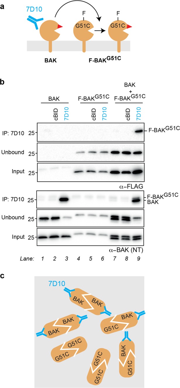
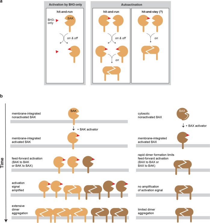
BAK and BAX, which drive commitment to apoptosis, are activated principally by certain BH3-only proteins that bind them and trigger major rearrangements. One crucial conformation change is exposure of their BH3 domain which allows BAK or BAX to form homodimers, and potentially to autoactivate other BAK and BAX molecules to ensure robust pore formation and cell death. Here, we test whether full-length BAK or mitochondrial BAX that are specifically activated by antibodies can then activate other BAK or BAX molecules. We found that antibody-activated BAK efficiently activated BAK as well as mitochondrial or cytosolic BAX, but antibody-activated BAX unexpectedly proved a poor activator. Notably, autoactivation by BAK involved transient interactions, as BAK and BAX molecules it activated could dissociate and homodimerize. The results suggest that BAK-driven autoactivation may play a substantial role in apoptosis, including recruitment of BAX to the mitochondria. Hence, directly targeting BAK rather than BAX may prove particularly effective in inhibiting unwanted apoptosis, or alternatively, inducing apoptosis in cancer cells.


Conflict of interest statement

The authors declare that they have no conflict of interest.

Figures

Fig. 1
Fig. 1. Schematic of BAK activation and autoactivation.
In healthy cells, nonactivated BAK is inserted into the mitochondrial outer membrane via its α9 transmembrane domain. Activation can be triggered by BH3-only proteins such as BID (by binding to the α2–α5 groove) or by antibody (binding to the N-terminus of the α1–α2 loop). The major conformation changes that occur during activation include separation of three components—the N-terminal α1–helix, the α2–α5 core, and the α6–α9 latch—leaving the protein with an exposed BH3 domain (α2; red triangle) and a modified α2–α5 hydrophobic groove. (BAK conformation change is also illustrated in an animation of autoactivation in Fig. S6.) Once activated, the BAK monomer can bind to other family members to form either homodimers that promote apoptosis, or heterodimers with a pro-survival relative (here MCL-1) that do not contribute to pore formation. The activated monomer may also activate (autoactivate) additional BAK (or BAX) molecules.
Fig. 2
Fig. 2. Antibody-activated BAK activates BAKG51C.
a Schematic of two BAK variants (BAK and F-BAKG51C) co-expressed in mitochondria and then incubated with the 7D10 antibody to directly activate BAK and expose its BH3 domain (red triangle). 7D10 cannot bind F-BAKG51C. b Co-immunoprecipitation shows that F-BAKG51C is activated by the 7D10 antibody if BAK is also present. Two BAK variants were expressed individually or together in Bax/−Bak−/− mouse embryonic fibroblasts (MEF), and membrane fractions incubated with the 7D10 antibody to activate (wild-type) BAK. As positive controls, aliquots were also incubated with cBID to activate both forms of BAK. Samples were immunoprecipitated using the 7D10 antibody, and immunoblotted for FLAG (upper panels) or BAK (lower panels). Data are representative of two independent experiments. c Schematic of BAK dimers captured by the 7D10 antibody. Activated BAK and activated F-BAKG51C associate to form homodimers, some of which 7D10 can immunoprecipitate.
Fig. 3
Fig. 3. Antibody-activated BAK can activate mitochondrial BAX.
a Schematic of BAK co-expressed with a mitochondrial form of BAX (BAX-S184L) and incubated with the 7D10 antibody to activate BAK. b Major proteinase K cleavage sites in activated BAK and BAX locate to the α1–α2 loop region. Fragment sizes detected by the 4B5 or clone 3 antibody (see Fig. 3c) are shown. Note that activated BAK is cleaved to a 17 kDa fragment (rather than a 15 kDa fragment) when 7D10 antibody is bound, because the antibody masks a cleavage site. c Conformation change in BAX-S184L is triggered by 7D10 antibody if BAK is also present. BAK and BAX-S184L were expressed individually or together in Bax−/−Bak−/− MEF, and membrane fractions incubated with the 7D10 antibody to activate BAK. As positive controls, aliquots were also incubated with cBID to activate both BAK and BAX-S184L. Samples were then incubated with proteinase K and western blotted to reveal fragments corresponding to nonactivated and activated forms of BAK (upper panel) or BAX-S184L (lower panel). Data are representative of two independent experiments. d Co-immunoprecipitation shows that BAX-S184L is activated by 7D10 antibody if BAK is also present. Membrane fractions were treated as in (c) and immunoprecipitated with 7D10, which had been added to the incubations to activate BAK. The samples were then immunoblotted for BAX-S184L (upper panels) or BAK (lower panels). Data are representative of two independent experiments. e Schematic of BAK and BAX-S184L dimers captured by the 7D10 antibody in panel (d). Activated BAK and activated BAX-S184L associate to form dimers, some of which 7D10 can immunoprecipitate.
Fig. 4
Fig. 4. Antibody-activated BAK at mitochondria recruits and activates wild-type BAX.
a Schematic of BAK combined with wild-type BAX and incubated with the 7D10 antibody to activate BAK. b Translocation and MOM insertion of recombinant BAX is triggered by 7D10 if BAK is also present. Bax−/−Bak−/− MEF or those cells expressing BAK were permeabilized and supplemented with 20 nM recombinant BAX, then incubated with 7D10 to activate BAK. As positive controls, aliquots were also incubated with cBID to activate both BAK and BAX. Samples were then centrifuged to recover the supernatant (Sup). The pellet fraction was then subjected to carbonate extraction to separate peripherally attached (Periph) and membrane-inserted (Insert) BAX. The three fractions were then immunoblotted for BAX. Data are representative of three independent experiments. c Translocation and activation of cytosolic BAX in permeabilized HeLa cells incubated with 7D10. HeLa cells were permeabilized and incubated with cBID, the 7D10 antibody to BAK, or the 3C10 antibody to BAX. The cytosolic and membrane fractions were then incubated with proteinase K and blotted for BAK (upper panel) or BAX (lower panel). Data are representative of two independent experiments. *Inhibitory effect of 3C10 on cytosolic BAX is evident from the inability of proteinase K to cleave α9. # Cross-reactive fragments from 7D10 and 3C10 antibodies.
Fig. 5
Fig. 5. Antibody-activated mitochondrial BAX-S184L is a poor activator of BAK, BAX-S184L, and BAX.
a Schematic of BAX-S184L co-expressed with BAK and incubated with 3C10 antibody, which directly activates BAX-S184L. b Conformation change in BAK is not triggered by 3C10 antibody, even if BAX-S184L is activated. BAX-S184L and BAK were expressed individually or together in Bax/−Bak−/− MEF, and membrane fractions incubated with the 3C10 antibody to activate BAX-S184L. As positive controls, aliquots were also incubated with cBID to activate both BAX-S184L and BAK. Samples were then incubated with proteinase K and western blotted to reveal fragments corresponding to nonactivated and activated forms of BAX-S184L (upper panel) or BAK (lower panel). Data are representative of two independent experiments. c Oligomerization of BAK is not triggered by 3C10 antibody, even if BAX-S184L has been activated and oligomerized. Aliquots from (b) were incubated with oxidant (CuPhe) to detect oligomerization. Note that BAX-S184L linkage at S55C:R94C detects BH3:groove homodimers (see schematic in Fig. S3), and that BAK linkage detects both C14:C166 linkage within nonactivated BAK (Mx) and linkage within and between dimers (2X). Data are representative of two independent experiments. d Schematic of F-BAX-S184L co-expressed with the BAX-S184LR34A variant and then incubated with 3C10 which directly activates BAX-S184L but not the R34A variant. e Immunoprecipitation shows that BAX-S184L is a poor activator of BAX-S184LR34A. BAX-S184L and BAX-S184LR34A were expressed individually or together in Bax−/−Bak/ MEF, and membrane fractions incubated with the 3C10 antibody to activate BAX-S184L. As positive controls, aliquots were also incubated with cBID to activate both BAX-S184L variants. The pellet fractions were then solubilized and an aliquot taken for the input sample. Additional aliquots were immunoprecipitated either with (upper panels) or without (lower panels) addition of the 6A7 antibody that binds to activated BAX, and the samples immunoblotted for BAX. Note that the beads used in the immunoprecipitation would bind both the 3C10 and 6A7 antibodies. Data are representative of two independent experiments. f Schematic of BAX-S184L combined with recombinant BAXR34A and incubated with the 3C10 antibody to directly activate BAX-S184L. 3C10 cannot bind the BAXR34A variant. g BAX-S184L is a poor activator of recombinant BAXR34A. Membrane fractions from Bax/−Bak/− MEF or those cells expressing BAX-S184L or BAK were supplemented with 50 nM recombinant BAXR34A and incubated with cBID or the 3C10 or 7D10 antibody. Samples were then subjected to carbonate extraction as in Fig. 4b and the supernatant (Sup.) and inserted (Insert.) fractions blotted for BAX. Note that F-BAX-S184L is partly peripherally attached, but becomes activated and inserted following either cBID or 3C10 treatment (lanes 5 and 6). Data are representative of three independent experiments.
Fig. 6
Fig. 6. Mutation in hydrophobic groove converts BAX into a strong activator.
a Schematic of recombinant BAXR109D incubated with BAK-expressing mouse liver mitochondria. b In the absence of an activating stimulus (e.g., no cBID or activating antibody), intermediate levels of BAX or BAXR109D can permeabilize Bak−/− mitochondria. Mouse liver mitochondria (MLM) from Bak−/− mice were incubated with increasing levels of recombinant BAX or the groove mutant BAXR109D and assessed for cytochrome c release. As positive controls, additional aliquots were incubated with 10 nM cBID and 10 nM BAX proteins. Data are representative of four independent experiments. c Activated BAXR109D but not wild-type BAX is able to autoactivate BAK. MLM from wild-type mice were incubated as in (b) and assessed for cytochrome c release (upper panels) and activation of mouse BAK (by proteinase K cleavage, lower panels). Data are representative of four independent experiments.
Fig. 7
Fig. 7. Summary.
a Schematic of the transient interaction involved in autoactivation by BAK. The α2–α5 hydrophobic groove activation site in BAK is used to illustrate the hit-and-run interaction involved in its activation by BH3-only proteins (left panel). Autoactivation also involves a transient interaction (middle panel) as the targeted BAKG51C and BAX-S184L proteins became nearly fully activated and could homodimerize rather than remain bound to BAK (see Figs. S2, 3c, and S3). Whether a proportion of targeted molecules can remain bound to BAK as a BH3:groove dimer (right panel; hit-and-stay) has not been determined. b The transient interaction involved in autoactivation by BAK (left panels) highlights the potential of directly targeting BAK to either inhibit or promote apoptosis, whereas direct BAX activation (right panels) is expected to be less productive.

References

    1. Bock, F. J. & Tait, S. W. G. Mitochondria as multifaceted regulators of cell death. Nat. Rev. Mol. Cell Biol. 10.1038/s41580-019-0173-8 (2019). - PubMed
    1. Moldoveanu, T. & Czabotar, P. E. BAX, BAK, and BOK: a coming of age for the BCL-2 family effector proteins. Cold Spring Harb. Perspect. Biol. 10.1101/cshperspect.a036319 (2019). - PMC - PubMed
    1. Westphal D, Kluck RM, Dewson G. Building blocks of the apoptotic pore: how Bax and Bak are activated and oligomerize during apoptosis. Cell Death Differ. 2014;21:196–205. doi: 10.1038/cdd.2013.139. - DOI - PMC - PubMed
    1. Chen L, et al. Differential targeting of prosurvival Bcl-2 proteins by their BH3-only ligands allows complementary apoptotic function. Mol. Cell. 2005;17:393–403. doi: 10.1016/j.molcel.2004.12.030. - DOI - PubMed
    1. Letai A, et al. Distinct BH3 domains either sensitize or activate mitochondrial apoptosis, serving as prototype cancer therapeutics. Cancer Cell. 2002;2:183–192. doi: 10.1016/S1535-6108(02)00127-7. - DOI - PubMed

Publication types

Substances