. 2020 Apr 3:2020:5076865.
doi: 10.1155/2020/5076865. eCollection 2020.

Influence of Patient-Specific Head Modeling on EEG Source Imaging

Affiliations

Influence of Patient-Specific Head Modeling on EEG Source Imaging

Yohan Céspedes-Villar et al. Comput Math Methods Med. .

Abstract

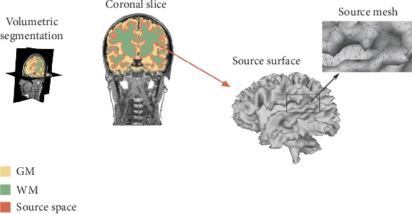
Electromagnetic source imaging (ESI) techniques have become one of the most common alternatives for understanding cognitive processes in the human brain and for guiding possible therapies for neurological diseases. However, ESI accuracy strongly depends on the forward model capabilities to accurately describe the subject's head anatomy from the available structural data. Attempting to improve the ESI performance, we enhance the brain structure model within the individual-defined forward problem formulation, combining the head geometry complexity of the modeled tissue compartments and the prior knowledge of the brain tissue morphology. We validate the proposed methodology using 25 subjects, from which a set of magnetic-resonance imaging scans is acquired, extracting the anatomical priors and an electroencephalography signal set needed for validating the ESI scenarios. Obtained results confirm that incorporating patient-specific head models enhances the performed accuracy and improves the localization of focal and deep sources.


Conflict of interest statement

The authors declare that there are no conflicts of interest regarding the publication of this paper.

Figures

Figure 1
Figure 1
Schematic methodology representation for enhancing the brain tissue model tested within the EEG forward problem formulation. The figure shows the proposed individually defined head modeling, including patient-dependent structural MRI, individual MRI segmentation, FDRM, and individual source space modeling. The middle and bottom boxes of (a) show the comparative structural information, namely, atlas (AT) and New York (NY). The top middle panel of (b) shows different tissue complexities (3L, 4L, and 5L). The remaining panels of (c) show the source localization and the performance measure.
Figure 2
Figure 2
Source space generation trough individually defined cortical meshes using morphological operators.
Figure 3
Figure 3
Exemplary of five-layered segmentation performed for the contrasted head models to incorporate prior information into the EEG forward model formulation.
Figure 4
Figure 4
Radar chart showing the ψ values achieved by both ESI solutions for each scenario and patient. The gray circle is not drawn in the LORETA chart since the difference of patients for each image data and configuration of tissue model complexity is much lower than 3 points.
Figure 5
Figure 5
Results of Bayesian model selection shown as the expected posterior probability and Bayesian omnibus risk assessed by each testing scenario of a brain tissue model. LORETA and MSP are the contrasted ESI solutions.
Figure 6
Figure 6
Achieved source reconstructions with NY models for both ESI methods. (a) Sensor space: ERP and topographic map. (b) Reconstructed activity. Views: Or: outside right; Ol: outside left; To: top; Bo: bottom; Ir: inside right; Il: inside left.
Figure 7
Figure 7
Results of group study shown as the expected posterior probability and Bayesian omnibus risk assessed by each testing scenario and MSP that is employed as ESI solution.
Figure 8
Figure 8
ESI solution for a representative subject using the best-achieved head models for each structural prior information, namely, 5L-PD, 3L-AT, and 5L-NY. (a) Sensor space: ERP and topographic map. (b) Reconstructed activity. Views: Or: outside right; Ol: outside left; To: top; Bo: bottom; Ir: inside right; Il: inside left.
Figure 9
Figure 9
Expected posterior probability and Bayesian omnibus risk for the group study of 3L and 4L scenarios for each case of image data.

References

    1. Grech R., Cassar T., Muscat J., et al. Review on solving the inverse problem in EEG source analysis. Journal of Neuroengineering and Rehabilitation. 2008;5(1):p. 25. doi: 10.1186/1743-0003-5-25. - DOI - PMC - PubMed
    1. Puce A., Hämäläinen M. A review of issues related to data acquisition and analysis in EEG/MEG studies. Brain Sciences. 2017;7(12):p. 58. doi: 10.3390/brainsci7060058. - DOI - PMC - PubMed
    1. Schlögl A., Keinrath C., Zimmermann D., Scherer R., Leeb R., Pfurtscheller G. A fully automated correction method of EOG artifacts in EEG recordings. Clinical Neurophysiology. 2007;118(1):98–104. doi: 10.1016/j.clinph.2006.09.003. - DOI - PubMed
    1. Delorme A., Sejnowski T., Makeig S. Enhanced detection of artifacts in EEG data using higher-order statistics and independent component analysis. NeuroImage. 2007;34(4):1443–1449. doi: 10.1016/j.neuroimage.2006.11.004. - DOI - PMC - PubMed
    1. Lepage K., Kramer M., Chu C. A statistically robust EEG re-referencing procedure to mitigate reference effect. Journal of Neuroscience Methods. 2014;235:101–116. doi: 10.1016/j.jneumeth.2014.05.008. - DOI - PMC - PubMed

Publication types

MeSH terms

LinkOut - more resources