Review
. 2018 Sep;110(5):e24040.
doi: 10.1002/pep2.24040. Epub 2018 Feb 13.

Membranotropic peptides mediating viral entry

Affiliations
Review

Membranotropic peptides mediating viral entry

Annarita Falanga et al. Pept Sci (Hoboken). 2018 Sep.

Abstract

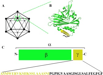
The means used by enveloped viruses to bypass cellular membranes are well characterized; however, the mechanisms used by non-enveloped viruses to deliver their genome inside the cell remain unresolved and poorly defined. The discovery of short, membrane interacting, amphipathic or hydrophobic sequences (known as membranotropic peptides) in both enveloped and non-enveloped viruses suggests that these small peptides are strongly involved in breaching the host membrane and in the delivery of the viral genome into the host cell. Thus, in spite of noticeable differences in entry, this short stretches of membranotropic peptides are probably associated with similar entry-related events. This review will uncover the intrinsic features of viral membranotropic peptides involved in viral entry of both naked viruses and the ones encircled with a biological membrane with the objective to better elucidate their different functional properties and possible applications in the biomedical field.

Keywords: enveloped viruses; fusion peptide; membranotropic peptides; non‐enveloped virus.


Figures

Figure 1
Figure 1
Different mechanisms of membrane interaction exploited for viral entry by non‐enveloped and enveloped viruses. Non‐enveloped viruses are reported on the left; capsid proteins release lytic factors responsible of nucleic acid internalization. Envelope viruses exploit the mechanism of membrane fusion either on the plasma membrane or within an endocytic vesicle
Figure 2
Figure 2
Schematic representation of the fusion process promoted by the three different classes of fusion proteins of enveloped viruses: Class I (panel A), Class II (panel B), and Class III (Panel C). Pre‐ and post‐fusion representation of fusion glycoproteins is reported. Fusion peptides are shown in red
Figure 3
Figure 3
Schematic representation of FHV capsid (A). An expanded view of the crystallographic structure (pdb: 4FTB) of one subunit (α protein) showing the location of the amphipathic region of γ peptide in yellow (B). Schematic representation of α protein, which undergoes auto cleavage during maturation producing β and γ (C) with relative sequence of γ peptide

References

    1. Yamauchi Y., Helenius A., J. Cell Sci. 2013, 126, 1289. - PubMed
    1. White J. M., Whittaker G. R., Traffic 2016, 17, 593. - PMC - PubMed
    1. Más V., Melero J. A., in Entry of Enveloped Viruses into Host Cells: Membrane Fusion (Ed: Mateu M. G.), Springer, Dordrecht, The Netherlands: 2013, 467. - PMC - PubMed
    1. Moyer C. L., Nemerow G. R., Curr. Opin. Virol. 2011, 1, 44. - PMC - PubMed
    1. Falanga A., Cantisani M., Pedone C., Galdiero S., Prot. Pept. Lett. 2009, 16, 751. - PubMed

LinkOut - more resources