

Cardiac Tamponade Secondary to COVID-19
- PMID: 32328588
- PMCID: PMC7177077
- DOI: 10.1016/j.jaccas.2020.04.009
Cardiac Tamponade Secondary to COVID-19
Abstract
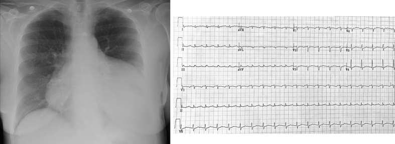
A 67-year-old woman presented with upper respiratory symptoms and was diagnosed with coronavirus disease-2019 (COVID-19). She was found to have a large hemorrhagic pericardial effusion with echocardiographic signs of tamponade and mild left ventricular impairment. Clinical course was complicated by development of takotsubo cardiomyopathy. She was treated with pericardiocentesis, colchicine, corticosteroids, and hydroxychloroquine, with improvement in symptoms. (Level of Difficulty: Intermediate.).
Keywords: COVID-19; COVID-19, coronavirus disease-2019; ECG, electrocardiography; LDH, lactate dehydrogenase; LVEF, left ventricular ejection fraction; RR, reference range; SARS-CoV-2, severe acute respiratory syndrome-coronavirus-2; TTC, takotsubo cardiomyopathy; TTE, transthoracic echocardiography; cTnI, cardiac troponin I; pericardial effusion; takotsubo cardiomyopathy; tamponade.
© 2020 The Authors.
Figures


References
-
- World Health Organization Naming the coronavirus disease (COVID-19) and the virus that causes it. https://www.who.int/emergencies/diseases/novel-coronavirus-2019/technica... Available at:
-
- Salehi S., Abedi A., Balakrishnan S., Gholamrezanezhad A. Coronavirus disease 2019 (COVID-19): a systematic review of imaging findings in 919 patients. AJR Am J Roentgenol. 2020;215:87–93. - PubMed
-
- Adler Y., Charron P., Imazio M. 2015 ESC Guidelines for the diagnosis and management of pericardial diseases: the Task Force for the Diagnosis and Management of Pericardial Diseases of the European Society of Cardiology (ESC) Endorsed by: The European Association for Cardio-Thoracic Surgery (EACTS) Eur Heart J. 2015;36:2921–2964. - PMC - PubMed
Publication types
LinkOut - more resources
Full Text Sources
Research Materials
Miscellaneous
