. 2020 Aug;56(4):448-460.
doi: 10.1007/s11262-020-01755-3. Epub 2020 Apr 23.

Isolation and characterization of new Puumala orthohantavirus strains from Germany

Affiliations

Isolation and characterization of new Puumala orthohantavirus strains from Germany

Florian Binder et al. Virus Genes. 2020 Aug.

Abstract

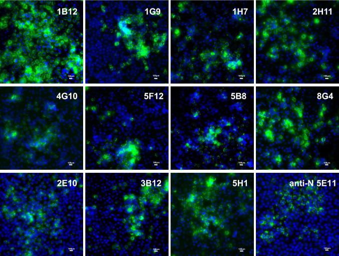
Orthohantaviruses are re-emerging rodent-borne pathogens distributed all over the world. Here, we report the isolation of a Puumala orthohantavirus (PUUV) strain from bank voles caught in a highly endemic region around the city Osnabrück, north-west Germany. Coding and non-coding sequences of all three segments (S, M, and L) were determined from original lung tissue, after isolation and after additional passaging in VeroE6 cells and a bank vole-derived kidney cell line. Different single amino acid substitutions were observed in the RNA-dependent RNA polymerase (RdRP) of the two stable PUUV isolates. The PUUV strain from VeroE6 cells showed a lower titer when propagated on bank vole cells compared to VeroE6 cells. Additionally, glycoprotein precursor (GPC)-derived virus-like particles of a German PUUV sequence allowed the generation of monoclonal antibodies that allowed the reliable detection of the isolated PUUV strain in the immunofluorescence assay. In conclusion, this is the first isolation of a PUUV strain from Central Europe and the generation of glycoprotein-specific monoclonal antibodies for this PUUV isolate. The obtained virus isolate and GPC-specific antibodies are instrumental tools for future reservoir host studies.

Keywords: Bank vole; Cell culture; Glycoprotein-specific antibodies; Puumala orthohantavirus; Virus adaptation.


Conflict of interest statement

The authors declare that they have no competing interests.

Figures

Fig. 1
Fig. 1
Schematic representation of the workflow. Bank voles were collected in forests within the district Osnabrück and a small piece of lung was taken by an incision in the chest area directly in the field. Lung tissue was meshed by grinding against a metal grid in a reaction tube containing 1 ml DMEM + 5% FCS and sterile filtered directly onto the cells. Cells were passaged three times until PUUV RT-qPCR screening. Supernatant of PUUV-positive flasks was taken and used for infection and further passaging in VeroE6 and MGN-2-R cells. Sequencing of complete genomes was done for PUUV RT-qPCR-positive passages and the corresponding original bank vole lung tissue. Isolates M52 and M62 were lost upon virus stock generation, presumably due to low viral load
Fig. 2
Fig. 2
Infection studies of PUUV isolates in VeroE6 and MGN-2-R cells. a Immunofluorescence analysis of VeroE6 and MGN-2-R cells inoculated with supernatants (SN) of PUUV Osnabrück/V29, isolated on VeroE6 cells, or PUUV Osnabrück/M43, isolated on MGN-2-R cells. PUUV-inoculated and mock-infected cells were fixed 10 days post infection and stained with nucleocapsid protein-specific antibody 5E11 and a secondary anti-mouse Alexa fluor 488 conjugated antibody. Nuclei were stained with DAPI. b Determination of virus titers of PUUV Osnabrück/V29 isolate (TCID50/ml) after three passages (P3) in VeroE6 and MGN-2-R cells (white columns) in comparison to titers directly after isolation in VeroE6 cells (black columns). Titers were obtained by immunofluorescence staining of 96-well plates 10 days post inoculation
Fig. 3
Fig. 3
Complete genome analysis of PUUV isolates. Complete PUUV genomes isolated from lung tissues and positive cell line passages were determined by HTS and dideoxy-chain termination sequencing in combination with RNA ligation to obtain complete NCRs. Nucleotide sequence insertion in the S segment NCR and amino acid exchanges in the L segment encoded RdRP are compared. Black dots indicate sequences derived from lung tissue and triangles indicate sequences obtained from cell culture passages. The complete genome of PUUV strain Astrup was used as a reference sequence ([21]; GenBank accession numbers: KJ994776-78). Coding regions of the three segments are indicated by numbers. NCR, non-coding region; M43, PUUV Osnabrück/MGN-2-R 43; V29, PUUV Osnabrück/VeroE6 29
Fig. 4
Fig. 4
Hantavirus phylogenetic trees. Hantavirus phylogenetic tree of concatenated S, M, and L coding sequences of 18 published and four novel complete genomes (a). Partial S segment coding sequences of 365 nucleotides length, reconstructed with four novel and 202 published partial sequences (b). New PUUV isolates (GenBank accession numbers: MN639737-MN639742) are indicated with triangles and sequences derived from their original lung tissue (GenBank accession numbers: MN639743-MN639748) are labeled with black dots. Published sequences of other hantaviruses are labeled with GenBank accession numbers. Novel sequences are highlighted in gray. Posterior probabilities for major nodes of the maximum clade credibility phylogenetic tree are displayed. Analysis was performed by Bayesian algorithms via MrBayes v.3.2.6 (https://sourceforge.net/projects/mrbayes/files/mrbayes/) on the CIPRES online portal [28]. A mixed nucleotide substitution matrix was specified in 4 independent runs of 107 generations. Scale bar indicates nucleotide substitutions per site. For clarity, previously characterized PUUV clades from other parts of Europe are shown in simplified form. CE, Central European; LAT, Latvian; ALAD, Alpe-Adrian; S-SCA, South Scandinavian; N-SCA, North Scandinavian; RUS, Russian; FIN, Finnish; DEN, Danish
Fig. 5
Fig. 5
Reactivity of novel PUUV GPC-specific monoclonal antibodies with hantavirus-infected VeroE6 cells in immunofluorescence assay (IFA). Antibodies were generated by immunization of BALB/c mice with GPC-derived virus-like particles of PUUV strain Astrup. After screening and subcloning, monoclonal antibodies were tested in IFA. VeroE6 cells were infected with PUUV Osnabrück V29 isolate on coverslips and fixed for IFA after 10 days. The monoclonal antibodies were administered for 1 h at RT. Detection of the specific antibody binding was done using an anti-mouse Alexa fluor 488 conjugated antibody. After staining, coverslips were mounted on glass slides for imaging

References

    1. Avsic-Zupanc T, Saksida A, Korva M. Hantavirus infections. Clin Microbiol Infect. 2015 doi: 10.1111/1469-0691.12291. - DOI - PubMed
    1. Krüger DH, Figueiredo LT, Song JW, Klempa B. Hantaviruses-globally emerging pathogens. J Clin Virol. 2015;64:128–136. doi: 10.1016/j.jcv.2014.08.033. - DOI - PubMed
    1. Faber M, Krüger DH, Auste B, Stark K, Hofmann J. Weiss S (2019) Molecular and epidemiological characteristics of human Puumala and Dobrava-Belgrade hantavirus infections, Germany, 2001 to 2017. Euro Surveillance. 2019 doi: 10.2807/1560-7917.Es.2019.24.32.1800675. - DOI - PMC - PubMed
    1. Reip A, Haring B, Sibold C, Stohwasser R, Bautz EK, Darai G, Meisel H, Kruger DH. Coding strategy of the S and M genomic segments of a hantavirus representing a new subtype of the Puumala serotype. Adv Virol. 1995;140(11):2011–2026. doi: 10.1007/bf01322689. - DOI - PubMed
    1. Elliott LH, Ksiazek TG, Rollin PE, Spiropoulou CF, Morzunov S, Monroe M, Goldsmith CS, Humphrey CD, Zaki SR, Krebs JW, et al. Isolation of the causative agent of hantavirus pulmonary syndrome. Am J Trop Med Hygiene. 1994;51(1):102–108. doi: 10.4269/ajtmh.1994.51.102. - DOI - PubMed

MeSH terms