. 2020 Nov;235(11):8520-8532.
doi: 10.1002/jcp.29696. Epub 2020 Apr 24.

Nuclear factor of activated T cells 1 and 2 are required for vertebral homeostasis

Affiliations

Nuclear factor of activated T cells 1 and 2 are required for vertebral homeostasis

Ernesto Canalis et al. J Cell Physiol. 2020 Nov.

Abstract

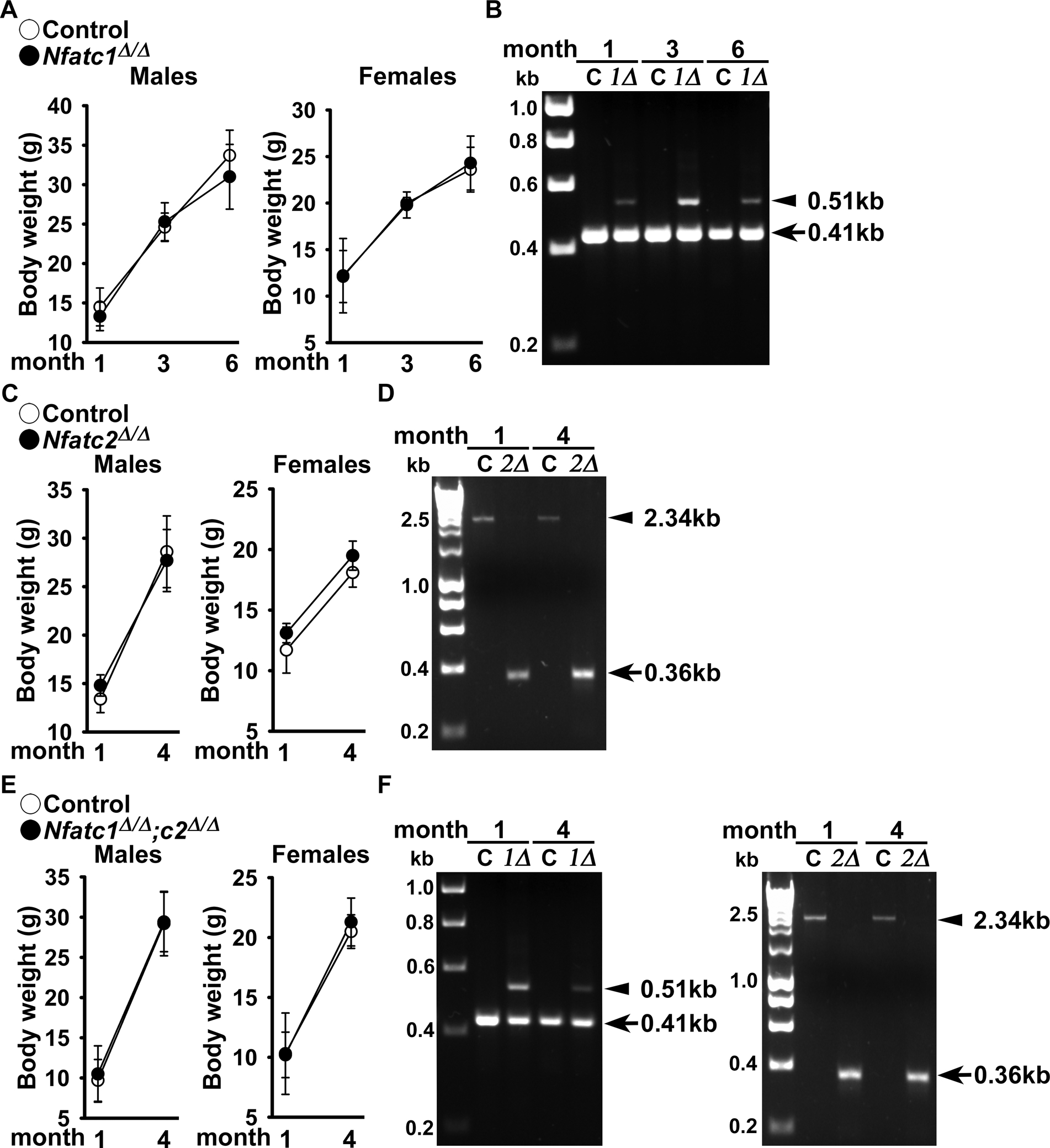
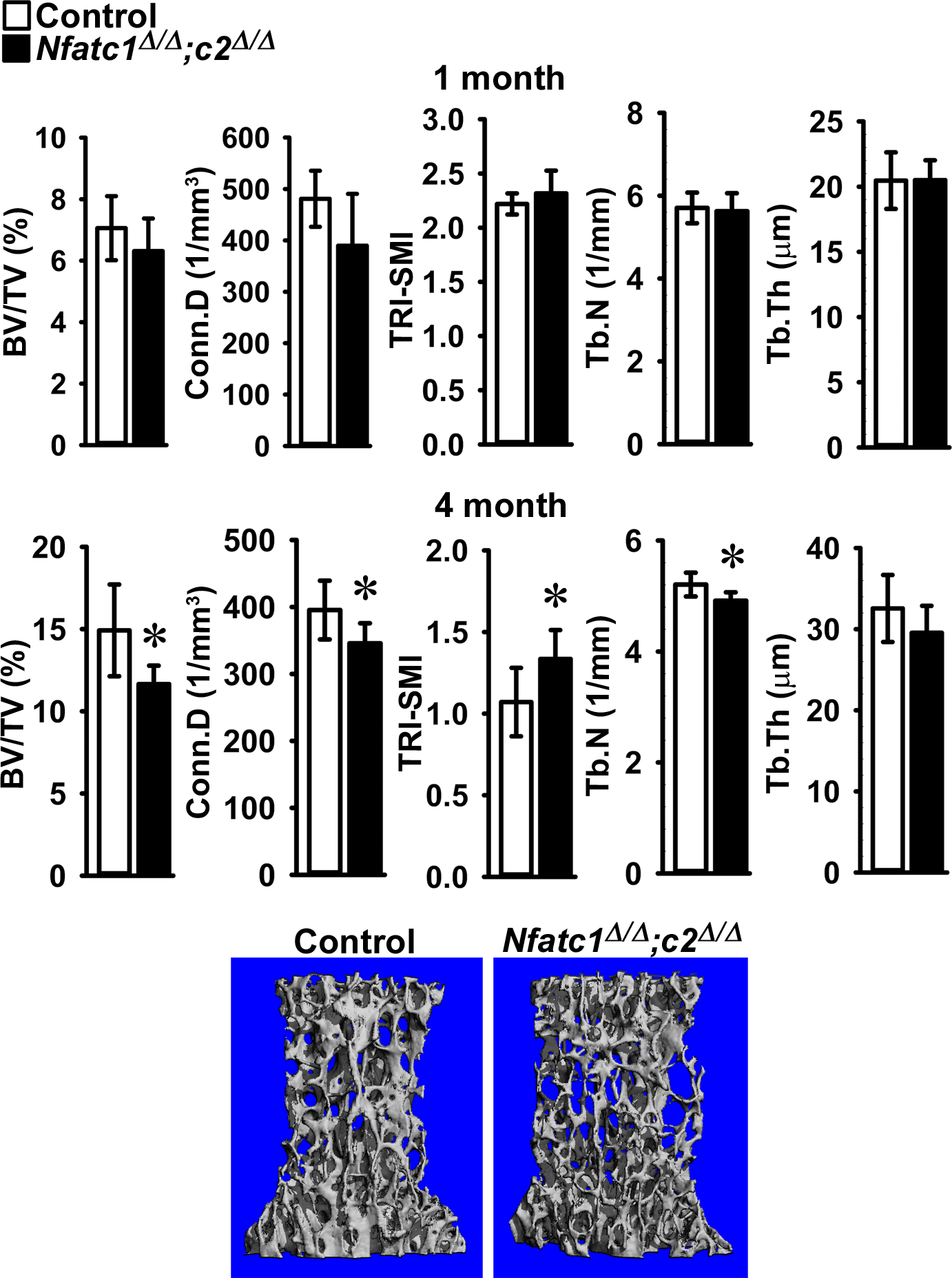
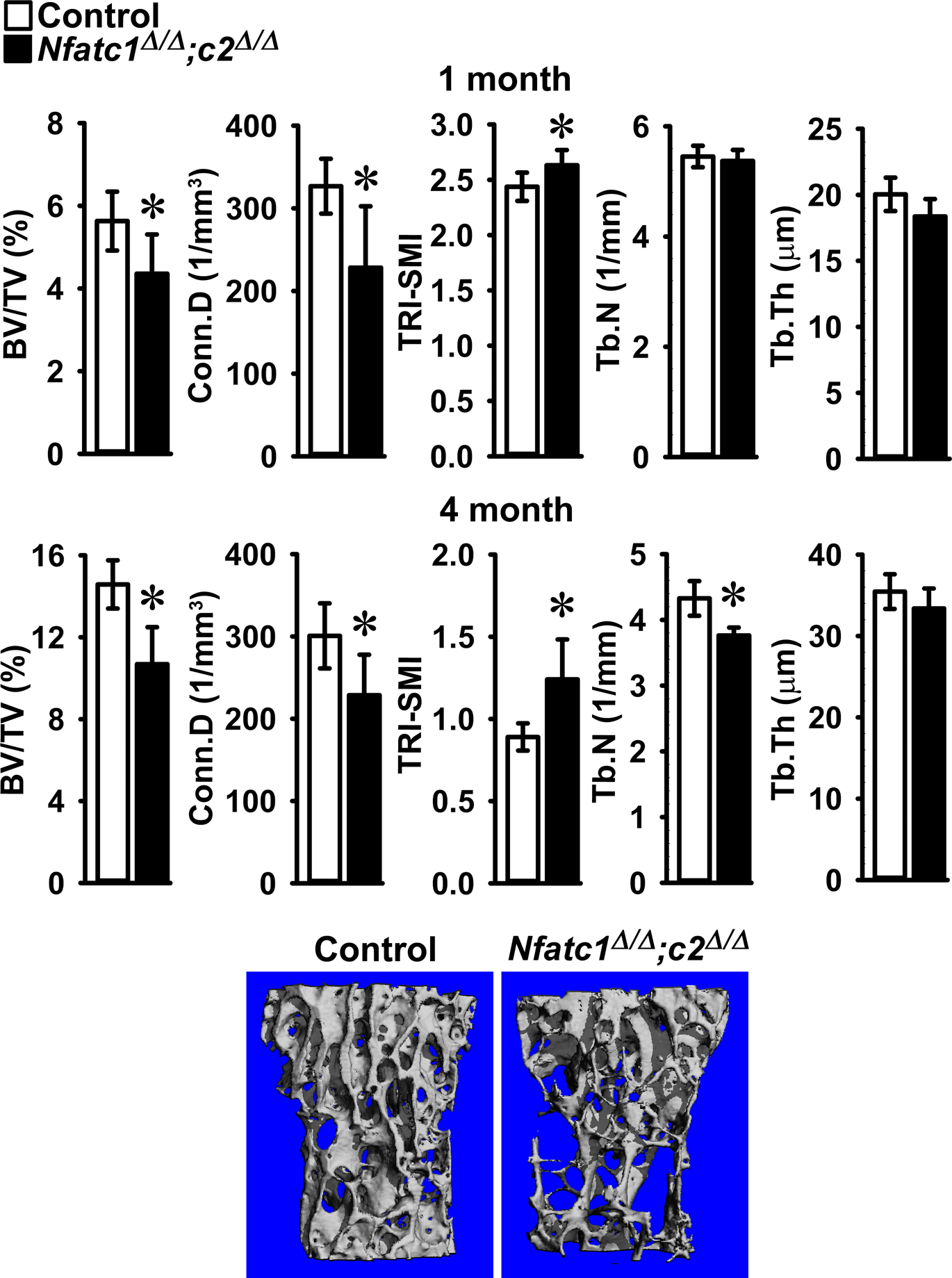
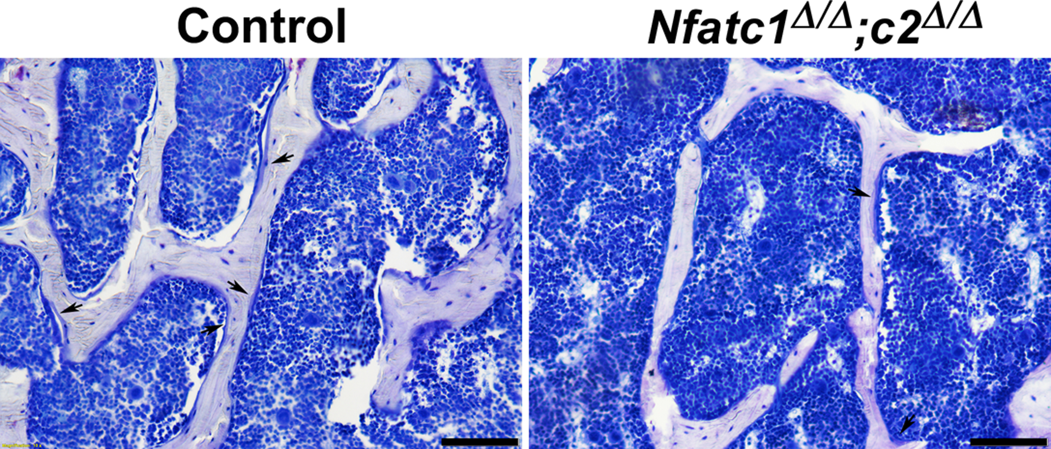
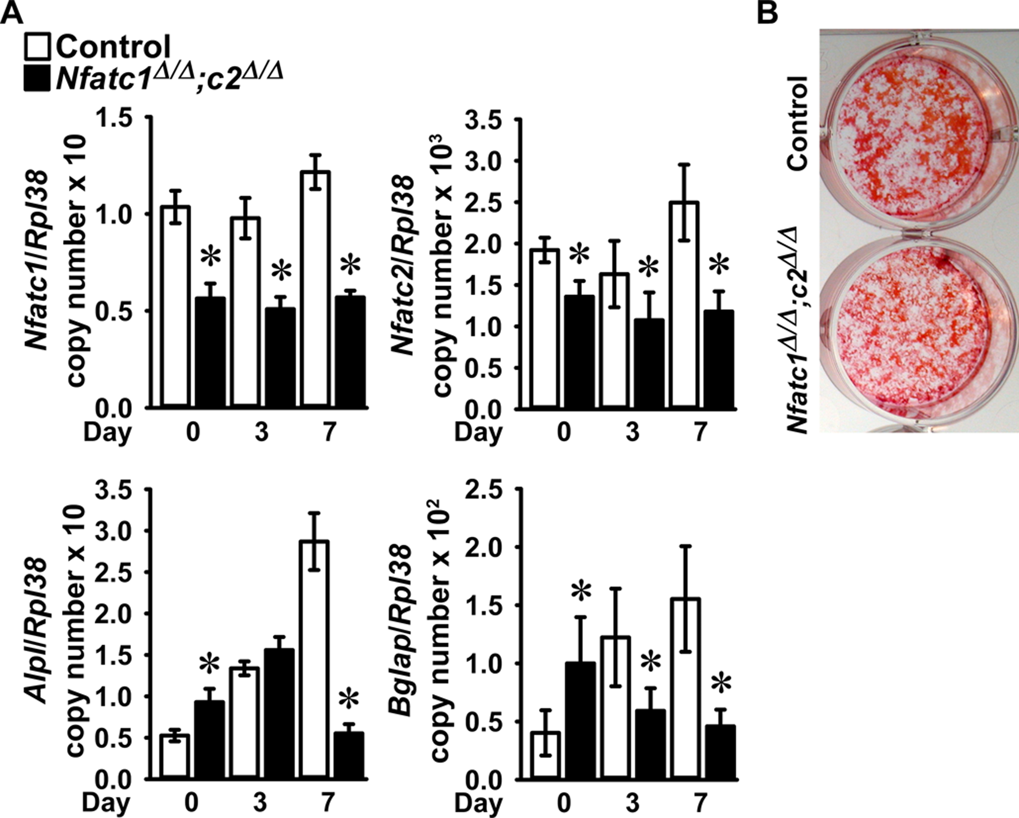
The present study defines the function of nuclear factor of activated T cells (NFAT)c1 and NFATc2 in osteoblast function in vivo and in vitro. Nfatc1loxP/loxP , Nfatc2loxP/loxP , and Nfatc1loxP/loxP ;Nfatc2loxP/loxP conditional mice were mated with BGLAP-Cre transgenics to inactivate Nfatc1 and Nfatc2 singly and in combination in osteoblasts. Microcomputed tomography demonstrated that male and female conditionally inactivated Nfatc1, Nfatc2 and dual Nfatc1;Nfatc2 mice had osteopenia at Lumbar 3 (L3) sites when compared to littermate controls. However, the Nfatc1 and Nfatc2 inactivation singly and in combination in Bglap-expressing osteoblasts did not result in an appreciable phenotype at femoral sites. Bone histomorphometry of L3 confirmed the osteopenic phenotype and demonstrated that Nfatc1;Nfatc2 inactivated male mice had a significant decrease in osteoblast number and in osteoblast surface and osteoid surface. The dual downregulation of Nfatc1 and Nfatc2 in bone marrow stromal cells caused a decrease in Alpl and Bglap expression, confirming a role of these transcription factors in osteoblast function. In conclusion, our studies reveal that NFATc1 and NFATc2 are necessary for optimal vertebral, but not femoral, bone homeostasis in vivo and osteoblast differentiation in vitro.

Keywords: NFATc2; bone formation; bone remodeling; osteoblasts.


Conflict of interest statement

Disclosures: The authors declare no conflicts of interest with the contents of this article. The data that support the findings of this study are available from the corresponding author upon reasonable request.

Figures

Figure 1.
Figure 1.
Weight and documentation of DNA recombination of Nfatc1 and Nfatc2 alleles by BGLAP-driven Cre. (A) Body weight of BGLAP-Cre;Nfatc1Δ/Δ, BGLAP-Cre;Nfatc2Δ/Δ and BGLAP-Cre;Nfatc1Δ/Δ;Nfatc2Δ/Δ and sex-matched control Nfatc1loxP/loxP, Nfatc2loxP/loxP and Nfatc1loxP/loxP;Nfatc2loxP/loxP littermates. Values are means ± SD; n = 3 control and n = 6 Nfatc1Δ/Δ; n = 5 control and n = 7 Nfatc2Δ/Δ and n = 3 control and n= 9 Nfatc1Δ/Δ;Nfatc2Δ/Δ. (B) DNA from tibiae were obtained from BGLAP-Cre;Nfatc1Δ/Δ and DNA from calvariae were obtained from BGLAP-Cre;Nfatc2Δ/Δ and from BGLAP-Cre;Nfatc1Δ/Δ;Nfatc2Δ/Δ neonates and from their respective (non-Cre) Nfatc1loxP/loxP, Nfatc2loxP/loxP and Nfatc1loxP/loxP;Nfatc2loxP/loxP controls. Cre-dependent DNA recombination was demonstrated by gel electrophoresis of PCR amplification products obtained with primers for the unrecombined Nfatc1loxP and Nfatc2loxP and for the Cre-mediated recombined Nfatc1Δ and Nfatc2Δ alleles. In Figure 1B and 1F left, the arrow heads indicate the position of the 410 base pair (bp) amplicon verifying the unrecombined Nfatc1loxP allele and 510 bp amplicon verifying the Cre-mediated recombined Nfatc1Δ allele. In Figure 1D and right, the arrow heads indicate the 2.3 kilobase (kb) amplicon verifying the unrecombined Nfatc2loxP allele, and the 0.36 kb amplicon verifying the Cre-mediated recombined Nfatc2Δ allele.
Figure 2.
Figure 2.
Cancellous bone microarchitecture assessed by μCT of vertebral body of Lumbar 3 (L3) from 1 (upper panels) and 4 (lower panels) month old BGLAP-Cre;Nfatc1Δ/Δ;Nfatc2Δ/Δ male mice (black bars) and sex-matched littermate Nfatc1loxP/loxP;Nfatc2loxP/loxP controls (white bars). Parameters shown are: bone volume/tissue volume (BV/TV); connectivity density (Conn.D); structure model index (SMI); trabecular number (Tb.N) and trabecular thickness (Tb,Th). Values are means ± SD; n = 5 to 6 for control and n = 6 to 9 for Nfatc1Δ/Δ;Nfatc2Δ/Δ mice. A representative image shows cancellous bone osteopenia in 4 month old BGLAP-Cre;Nfatc1Δ/Δ;Nfatc2Δ/Δ male mice.
Figure 3.
Figure 3.
Cancellous bone microarchitecture assessed by μCT of vertebral body of Lumbar 3 (L3) from 1 (upper panels) and 4 (lower panels) month old BGLAP-Cre;Nfatc1Δ/Δ;Nfatc2Δ/Δ female mice (black bars) and sex-matched littermate Nfatc1loxP/loxP;Nfatc2loxP/loxP controls (white bars). Parameters shown are: bone volume/tissue volume (BV/TV); connectivity density (Conn.D); structure model index (SMI); trabecular number (Tb.N) and trabecular thickness (Tb.Th). Values are means ± SD; n = 5 for control and n = 8 for Nfatc1Δ/Δ;Nfatc2Δ/Δ mice. A representative image shows cancellous bone osteopenia in 4 month old BGLAP-Cre;Nfatc1Δ/Δ;Nfatc2Δ/Δ female mice.
Figure 4.
Figure 4.
Representative image of bone histomorphometry from 4 month old BGLAP-Cre;Nfatc1Δ/Δ;Nfatc2Δ/Δ male mice and control littermates. Sections were stained with toluidine blue. Arrows point to osteoblasts on osteoid surfaces and bar in right corner represents 100 μm.
Figure 5.
Figure 5.
Bone marrow stromal cells harvested from femurs of 6 to 8 week old male Nfatc1loxP/loxP;Nfatc2loxP/loxP mice were cultured to subconfluence, transduced with Ad-CMV-Cre (Nfatc1Δ/Δ;Nfatc2Δ/Δ, black bars) or Ad-CMV-GFP (control, white bars) and cultured under conditions favoring osteoblastogenesis following confluence (Day 0). In Panel A, total RNA was extracted and gene expression determined by qRT-PCR. Data are expressed as Nfatc1, Nfatc2, Alpl and Bglap copy number corrected for Rpl38 copy number. Values are means ± SD; n = 4. *Significantly different between Nfatc1Δ/Δ;Nfatc2Δ/Δ and control, p < 0.05. In Panel B, a representative image shows control and Nfatc1Δ/Δ;Nfatc2Δ/Δ 14 day cultures stained with alizarin red.

References

    1. Aliprantis AO, Ueki Y, Sulyanto R, Park A, Sigrist KS, Sharma SM, … Glimcher LH (2008). NFATc1 in mice represses osteoprotegerin during osteoclastogenesis and dissociates systemic osteopenia from inflammation in cherubism. The Journal of clinical investigation, 118(11), 3775–3789. - PMC - PubMed
    1. Asagiri M, Sato K, Usami T, Ochi S, Nishina H, Yoshida H, … Takayanagi H (2005). Autoamplification of NFATc1 expression determines its essential role in bone homeostasis. Journal of Experimental Medicine, 202(9), 1261–1269. - PMC - PubMed
    1. Bauer W, Rauner M, Haase M, Kujawski S, Arabanian LS, Habermann I, … Kiani A (2011). Osteomyelosclerosis, anemia and extramedullary hematopoiesis in mice lacking the transcription factor NFATc2. Haematologica, 96(11), 1580–1588. doi:10.3324/haematol.2011.042515 - DOI - PMC - PubMed
    1. Bianco P, & Gehron RP (2000). Marrow stromal stem cells. The Journal of clinical investigation, 105(12), 1663–1668. - PMC - PubMed
    1. Bouxsein ML, Boyd SK, Christiansen BA, Guldberg RE, Jepsen KJ, & Muller R (2010). Guidelines for assessment of bone microstructure in rodents using micro-computed tomography. Journal of Bone and Mineral Research, 25(7), 1468–1486. - PubMed

Publication types

MeSH terms

Substances