Review
. 2021 Jan;34(1):59-71.
doi: 10.1111/pcmr.12885. Epub 2020 Jun 17.

Acral lentiginous melanoma: Basic facts, biological characteristics and research perspectives of an understudied disease

Affiliations
Review

Acral lentiginous melanoma: Basic facts, biological characteristics and research perspectives of an understudied disease

Patricia Basurto-Lozada et al. Pigment Cell Melanoma Res. 2021 Jan.

Abstract

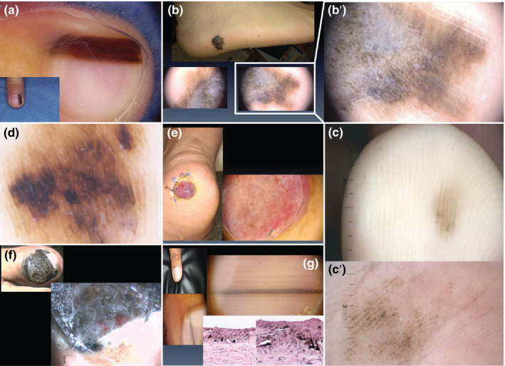
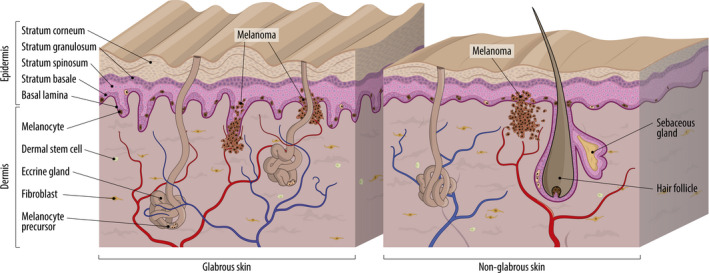
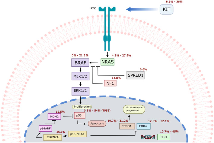
Acral lentiginous melanoma is a histological subtype of cutaneous melanoma that occurs in the glabrous skin of the palms, soles and the nail unit. Although in some countries, particularly in Latin America, Africa and Asia, it represents the most frequently diagnosed subtype of the disease, it only represents a small proportion of melanoma cases in European-descent populations, which is partially why it has not been studied to the same extent as other forms of melanoma. As a result, its unique genomic drivers remain comparatively poorly explored, as well as its causes, with current evidence supporting a UV-independent path to tumorigenesis. In this review, we discuss current knowledge of the aetiology and diagnostic criteria of acral lentiginous melanoma, as well as its epidemiological and histopathological characteristics. We also describe what is known about the genomic landscape of this disease and review the available biological models to explore potential therapeutic targets.

Keywords: acral melanoma; diagnosis; epidemiology; genomics; microenvironment.


Conflict of interest statement

None declared.

Figures

FIGURE 1
FIGURE 1
Examples of distinct acral lesions. (a) Longitudinal melanonychia in a 6‐year‐old patient with a subungual naevus. (b) Clinical appearance of an acral lentiginous melanoma (top panel), for which dermoscopy shows a parallel ridge pattern (bottom panels). (b′) Close‐up of the parallel ridge pattern typical of acral lentiginous melanoma. (c) Clinical appearance of an acral naevus showing a parallel furrow pattern. (c′) Close‐up of the parallel furrow pattern typical of acral naevi. (d) Dermoscopic appearance of a diffuse irregular pigmentation pattern in an advanced‐stage acral lentiginous melanoma. (e) Clinical and dermoscopic appearance of an amelanotic nodular melanoma on the heel. (f) Advanced‐stage nail unit melanoma. Dermoscopy is not necessary to make a diagnosis in these cases. (g) Adult patient with new onset of a pigmented lesion on the nail unit; clinically, a longitudinal melanonychia affecting the first finger. The recent onset, the affection of the first finger and the grey coloration shown by dermoscopy were important clues to suggest this was a nail unit melanoma despite the fact that there was no loss of parallelism nor affection >2/3 of the nail plate. All patients depicted here signed an informed consent that authorized the use of their photographs for research purposes
FIGURE 2
FIGURE 2
Schematic representation of glabrous and non‐glabrous skin. A thick stratum corneum, a grooved surface alternating ridges and sulci and the absence of hair follicles are some of the characteristics that define glabrous skin. Melanocytes are located in the basal layer of the epidermis of both skin types. Melanocyte precursors have been detected in hair follicles of non‐glabrous skin and in eccrine glands of glabrous skin. Evidence suggests that the epidermis may be a source of melanocyte precursors independent of skin appendages. Dermal stem cells can also differentiate into melanocytes. In glabrous skin, oncogenic mutations in melanocyte precursors have been observed in eccrine glands, suggesting that these can be transformed into melanoma cells. Other characteristics specific of glabrous skin such as the presence of encapsulated sense organs are not illustrated in the figure
FIGURE 3
FIGURE 3
Representation of main pathways affected in acral lentiginous melanoma. Proportions of ALM cases that harbour alterations in the genes depicted are shown in red, based on the available data (Puig‐Butillé et al., 2013; Rabbie, Ferguson, Molina‐Aguilar, Adams, & Robles‐Espinoza, 2019; Shim et al., 2017; Yeh et al., 2019)

References

    1. Albreski, D. , & Sloan, S. B. (2009). Melanoma of the feet: Misdiagnosed and misunderstood. Clinics in Dermatology, 27(6), 556–563. 10.1016/j.clindermatol.2008.09.014 - DOI - PubMed
    1. Alexandrescu, D. T. (2009). Melanoma costs: A dynamic model comparing estimated overall costs of various clinical stages. Dermatology Online Journal, 15(11), 1. - PubMed
    1. Alexandrov, L. B. , Nik‐Zainal, S. , Wedge, D. C. , Aparicio, S. A. J. R. , Behjati, S. , Biankin, A. V. , … Stratton, M. R. (2013). Signatures of mutational processes in human cancer. Nature, 500(7463), 415–421. - PMC - PubMed
    1. Al‐Hassani, F. , Chang, C. , & Peach, H. (2017). Acral lentiginous melanoma – Is inflammation the missing link? JPRAS Open, 14, 49–54. 10.1016/j.jpra.2017.06.002 - DOI
    1. André, J. , & Lateur, N. (2006). Pigmented nail disorders. Dermatologic Clinics, 24(3), 329–339. 10.1016/j.det.2006.03.012 - DOI - PubMed

Publication types