

PET-FDG: Impetus
- PMID: 32331374
- PMCID: PMC7226158
- DOI: 10.3390/cancers12041030
PET-FDG: Impetus
Abstract
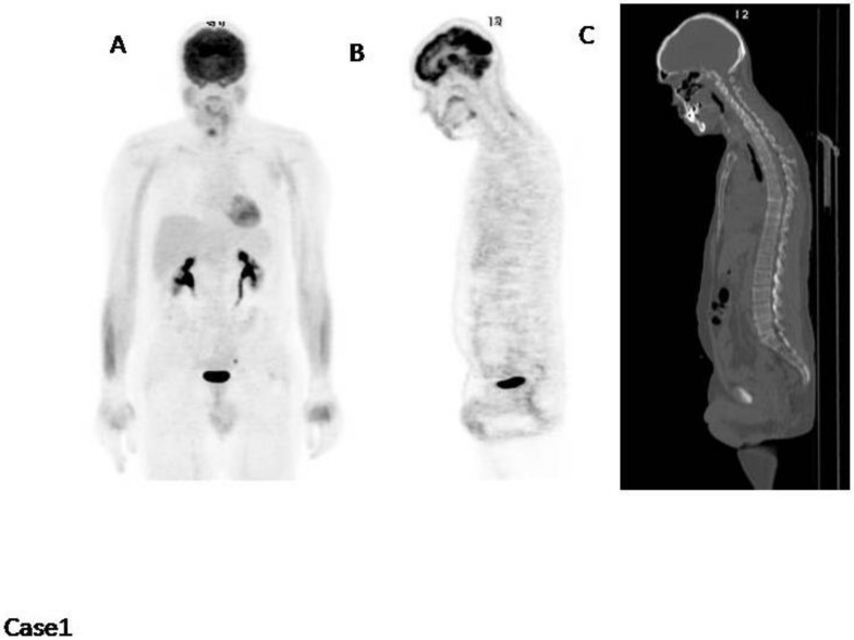
The International Myeloma Working Group (IMWG)recommends FDG PET/CT (Fluoro-Deoxy-glucose Positron Emission Tomography/Computed Tomography) as the gold standard imaging modality for initial evaluation and response to therapy assessment in multiple myeloma. In fact, FDG PET/CT, provides multiple useful indexes to risk-stratify patients and has significant prognostic value. However, multiple myeloma remains a complex disease to interpret on imaging. The Italian myeloma criteria for PET use (IMPeTUs) were proposed to standardize FDG PET/CT reading in multiple myeloma. In this communication an overview on IMPeTUs is provided as well as some examples of application.
Keywords: FDG; IMPeTUs; PET; interpretation criteria; multiple myeloma.
Conflict of interest statement
The authors declare no conflict of interest.
Figures


References
-
- Rajkumar S.V., Dimopolous M.A., Palumbo A., Blade J., Merlini G., Mateos M.V., Kumar S., Hillengass J., Kastritis E., Richardson P., et al. International Myeloma Working Group updated criteria for the diagnosis of multiple myeloma. Lancet Oncol. 2014;15:538–548. doi: 10.1016/S1470-2045(14)70442-5. - DOI - PubMed
-
- Rasche L., Angtuaco E., McDonald J.E., Buros A., Stein C., Pawlyn C., Thanendrarajan S., Schinke C., Samant R., Yaccoby S., et al. Low expression of hexokinase-2 is associated with false-negative FDG-positron emission tomography in multiple myeloma. Blood. 2017;130:30–34. doi: 10.1182/blood-2017-03-774422. - DOI - PMC - PubMed
-
- Abe Y., Ikeda S., Kitadate A., Narita K., Kobayashi H., Miura D., Takeuchi M., O’uchi E., O’uchi T., Matsue K. Low hexokinase-2 expression-associated false-negative 18F-FDG PET/CT as a potential prognostic predictor in patients with multiple myeloma. Eur. J. Nucl. Med. Mol. Imaging. 2019;46:1345–1350. doi: 10.1007/s00259-019-04312-9. - DOI - PubMed
-
- Dimopoulos M.A., Terpos E., Chanan-Khan A., Leung N., Ludwig H., Jagannath S., Niesvizky R., Giralt S., Fermand J.P., Bladé J., et al. Renal impairment in patients with multiple myeloma: A consensus statement on behalf of the International Myeloma Working Group. J. Clin. Oncol. 2010;28:4976–4984. doi: 10.1200/JCO.2010.30.8791. - DOI - PubMed
LinkOut - more resources
Full Text Sources
