Review
. 2020 Apr 22;10(4):244.
doi: 10.3390/diagnostics10040244.

HRCT Patterns of Drug-Induced Interstitial Lung Diseases: A Review

Affiliations
Review

HRCT Patterns of Drug-Induced Interstitial Lung Diseases: A Review

Giulio Distefano et al. Diagnostics (Basel). .

Abstract

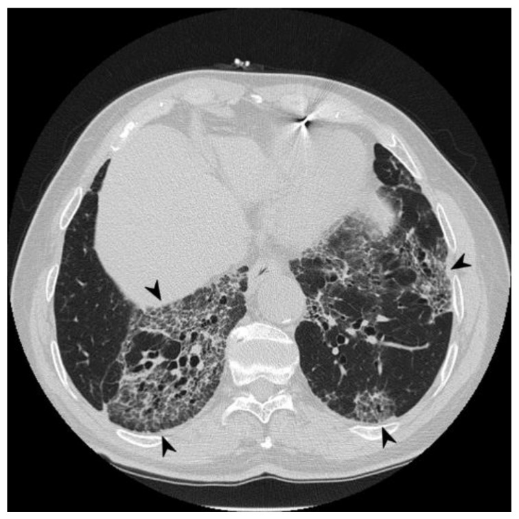
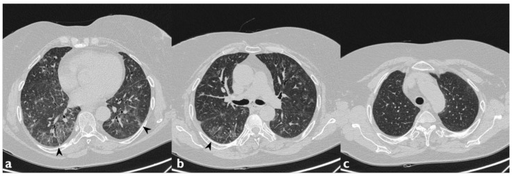
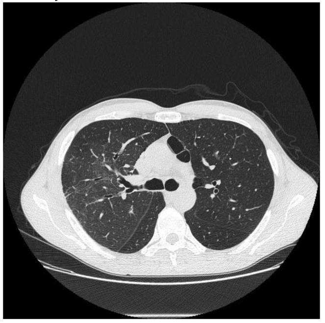
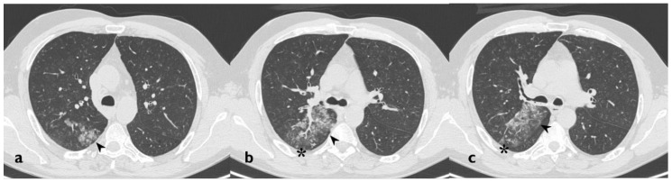
Interstitial Lung Diseases (ILDs) represent a heterogeneous group of pathologies, which may be related to different causes. A low percentage of these lung diseases may be secondary to the administration of drugs or substances. Through the PubMed database, an extensive search was performed in the fields of drug toxicity and interstitial lung disease. We have evaluated the different classes of drugs associated with pulmonary toxicity. Several different high resolution computed tomography (HRCT) patterns related to pulmonary drug toxicity have been reported in literature, and the most frequent ILDs patterns reported include Nonspecific Interstitial Pneumonia (NSIP), Usual Interstitial Pneumonia (UIP), Hypersensitivity Pneumonitis (HP), Organizing Pneumonia (OP), Acute Respiratory Distress Syndrome (ARDS), and Diffuse Alveolar Damage (DAD). Finally, from the electronic database of our Institute we have selected and commented on some cases of drug-induced lung diseases related to the administration of common drugs. As the imaging patterns are rarely specific, an accurate evaluation of the clinical history is required and a multidisciplinary approach-involving pneumologists, cardiologists, radiologists, pathologists, and rheumatologists-is recommended.

Keywords: acute; idiopathic pulmonary fibrosis; interstitial; lung diseases; multidetector computed tomography; respiratory distress syndrome; toxicity.


Conflict of interest statement

The authors declare no conflicts of interest.

Figures

Figure 1
Figure 1
A patient with methotrexate induced lung toxicity. Axial scan passing through the bases (a), through the origin of the pulmonary artery (b), and through the apices (c). At the level of the lower lobes there are multiple areas of ground glass opacity. In this case, interlobular septa thickening and initial signs of lung architectural distortion are also evident: these findings are not commonly encountered in patients with methotrexate toxicity, but they have been also reported in literature. Therefore, they may represent a possible trap in the diagnosis.
Figure 2
Figure 2
A case of suspected tocilizumab-induced lung toxicity. Axial scan passing through the bases (a), through the origin of the pulmonary artery, (b) and through the apices (c). Multiple areas of Ground-Glass Opacity (GGO), partly tending to confluence, predominantly located in the central regions of the lungs, partial sparing subpleural areas; fibrotic and nonspecific linear opacities are also shown in right lower lobe.
Figure 3
Figure 3
Cyclophosphamide-induced toxicity. Axial scan passing through the bases (a), through the origin of the pulmonary artery (b), and through the apices (c). Parenchymal consolidations are clearly recognizable in the upper lobes (black arrowheads); it is also possible to appreciate shaded areas of increased attenuation of the lung parenchyma as GGO spread to all segments (asterisk). Lung bases are less involved, as clearly depicted in figure a.
Figure 4
Figure 4
A case of Amiodarone-Induced Lung Toxicity (AILT). Axial scan passing through the bases (a), through the origin of the pulmonary artery (b), and through the apices (c). Reticulations, traction bronchiectasis, and widespread areas of GGO are shown in panels a–c (black arrowheads); parenchymal alterations have central and peripheral distribution. At the bases, in the subpleural field, an initial honeycomb pattern is appreciable (asterisk).
Figure 5
Figure 5
Another case of AILT. Axial scan passing through the bases (a), through the origin of the pulmonary artery (b), and through the apices (c). Reticular interstitial pattern superimposed to areas of GGO, distributed mainly to the lower lobes bilaterally and at lingula (black arrowheads); multiple traction bronchiectasis and bronchioloectasie are also present.
Figure 6
Figure 6
Same patient as the previous figure, follow-up two years after discontinuation of therapy: the scans passing through the basal segments demonstrate the substantial stability of the radiological picture (black arrowheads indicate the previous findings).
Figure 7
Figure 7
Another case of AILT. Axial scan passing through the bases (a), through the origin of the pulmonary artery (b), and through the apices (c). Diffuse reticular interstitial pattern and GGO, with multiple bronchiectasis and subpleural consolidation areas; these morphological features resemble a Nonspecific Interstitial Pneumonia (NSIP) pattern secondary to drug toxicity.
Figure 8
Figure 8
Another case of AILT. Axial scan passing through the bases (a), through the origin of the pulmonary artery (b), and through the apices (c). Interstitial disease with NSIP pattern. Diffuse increase in density of the lung parenchyma with a GGO appearance (black arrowheads indicate the previous findings).
Figure 9
Figure 9
Lung cocaine toxicity in a patient admitted to the emergency department with hemoptysis and dyspnea 24 h after inhalation of cocaine. Focal area of GGO, smooth septal thickening, and centrilobular nodule are visible in the right upper lobe.
Figure 10
Figure 10
Another case of lung cocaine toxicity. Axial scan passing through the upper lobes (ac). Focal area of GGO (black arrowheads), centrilobular nodule, and the tree-in-bud pattern (asterisk) are visible in the right upper lobe.
Figure 11
Figure 11
Association between HRCT patterns and the drugs most frequently responsible for lung toxicity.

References

    1. Cleverley J.R., Screaton N.J., Hiorns M.P., Flint J.D., Müller N.L. Drug-induced lung disease: High-resolution CT and histological findings. Clin. Radiol. 2002;57:292–299. doi: 10.1053/crad.2001.0792. - DOI - PubMed
    1. Lateef O., Shakoor N., Balk R.A. Methotrexate pulmonary toxicity. Expert Opin. Drug Saf. 2005;4:723–730. doi: 10.1517/14740338.4.4.723. - DOI - PubMed
    1. Edwards I.R., Aronson J.K. Adverse drug reactions: Definitions, diagnosis, and management. Lancet. 2000;356:1255–1259. doi: 10.1016/S0140-6736(00)02799-9. - DOI - PubMed
    1. Pirmohamed M., James S., Meakin S., Green C., Scott A.K., Walley T.J., Farrar K., Park B.K., Breckenridge A.M. Adverse drug reactions as cause of admission to hospital: Prospective analysis of 18,820 patients. BMJ. 2004;329:15–19. doi: 10.1136/bmj.329.7456.15. - DOI - PMC - PubMed
    1. Farcas A., Sinpetrean A., Mogosan C., Palage M., Vostinaru O., Bolita M., Domitrascu D. Adverse drug reactions detected by stimulated spontaneous reporting in an internal medicine department in Romania. Eur. J. Intern. Med. 2010;21:453–457. doi: 10.1016/j.ejim.2010.05.014. - DOI - PubMed