. 2020 Apr 22;25(8):1949.
doi: 10.3390/molecules25081949.

Biodegradable Cell Microcarriers Based on Chitosan/Polyester Graft-Copolymers

Affiliations

Biodegradable Cell Microcarriers Based on Chitosan/Polyester Graft-Copolymers

Tatiana S Demina et al. Molecules. .

Abstract

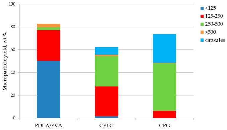
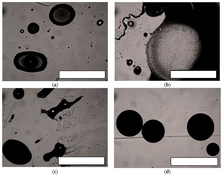
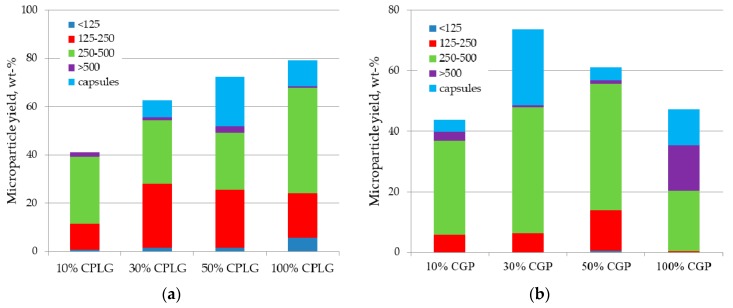
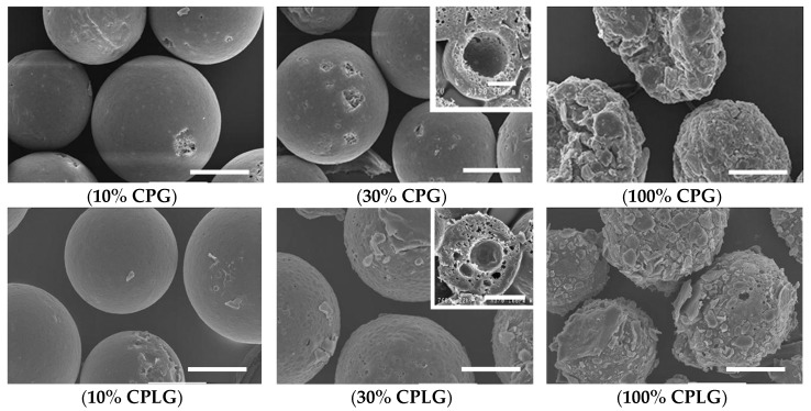
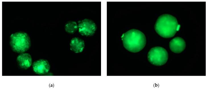
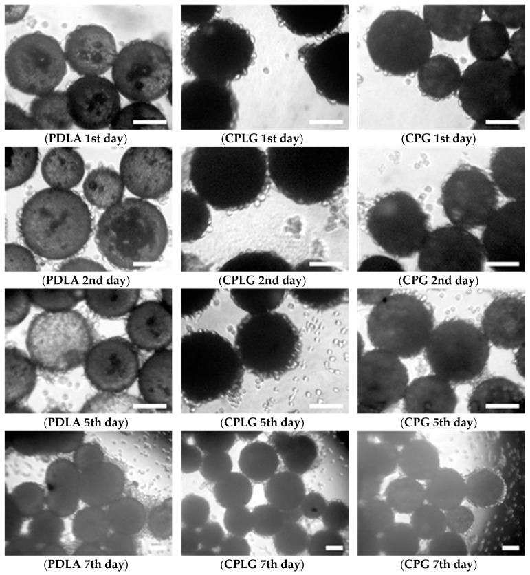
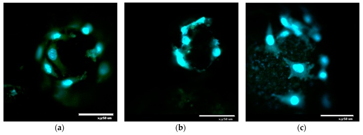
Self-stabilizing biodegradable microcarriers were produced via an oil/water solvent evaporation technique using amphiphilic chitosan-g-polyester copolymers as a core material in oil phase without the addition of any emulsifier in aqueous phase. The total yield of the copolymer-based microparticles reached up to 79 wt. %, which is comparable to a yield achievable using traditional emulsifiers. The kinetics of microparticle self-stabilization, monitored during their process, were correlated to the migration of hydrophilic copolymer's moieties to the oil/water interface. With a favorable surface/volume ratio and the presence of bioadhesive natural fragments anchored to their surface, the performance of these novel microcarriers has been highlighted by evaluating cell morphology and proliferation within a week of cell cultivation in vitro.

Keywords: chitosan; fibroblasts; graft-copolymers; microcarriers; oil/water emulsion; polylactide; tissue engineering.


Conflict of interest statement

The authors declare no conflict of interest.

Figures

Figure 1
Figure 1
Effect of 30 wt. % replacement of PDLA in oil phase by CPG or CPLG copolymers on the processing yield and size distribution of the microspheres and capsule formation.
Figure 2
Figure 2
Micrographies acquired under optical transmission microscopes of aliquots of the oil/water emulsion containing CPG (10 wt. %)/PDLA in the oil phase and withdrawn at (a) 5 min, (b) 15 min, (c) 25 min and (d) 35 min after the dispersion onset. Scale bar is 1000 μm.
Figure 3
Figure 3
Effect of (a) CPLG or (b) CPG copolymers content in oil phase on the processing yield and size distribution of the microspheres.
Figure 4
Figure 4
SEM micrographies of microparticles obtained by modifying the composition of the copolymer and its relative content in the oil phase of the o/w dispersion. The wt. % refers to the copolymer content to the total material in oil phase. Scale bars: 100 μm.
Figure 5
Figure 5
Micrographies of FITC-stained microparticles made from 30 wt. % of CPLG (a) and CPG (b) observed under fluorescence microscopy (Ex/Em 495/519 nm, respectively).
Figure 6
Figure 6
Microphotographies of L929 fibroblasts cultured on CPLG, CPG and PDLA microparticles. Scale bar is 100 μm.
Figure 7
Figure 7
Confocal microphotographies of L929 fibroblasts after 3 days of culture on (a) control PDLA, 30 wt. % (b) CPLG- and (c) CPG-contained microparticles. Scale bar is 50 μm. Living cells were stained with vital fluorescent dye Calcein AM and cell nuclei were stained with DAPI.

References

    1. Van Wezel A.L. Growth of Cell-Strains and Primary Cells on Micro-Carriers in Homogeneous Culture. Nature. 1967;216:64–65. doi: 10.1038/216064a0. - DOI - PubMed
    1. Markvicheva E., Grandfils C. Microcarriers for Animal Cell Culture. In: Nedovic V., Willaert R., editors. Fundamentals of Cell Immobilisation Biotechnology. Kluwer Academic Publishers; Berlin, Germany: 2004. pp. 141–161.
    1. Chen A.K.-L., Chen X., Choo A.B.H., Reuveny S., Oh S.K.W. Critical Microcarrier Properties Affecting the Expansion of Undifferentiated Human Embryonic Stem Cells. Stem Cell Res. 2011;7:97–111. doi: 10.1016/j.scr.2011.04.007. - DOI - PubMed
    1. Baakdhah T., van der Kooy D. Expansion of Retinal Stem Cells and Their Progeny Using Cell Microcarriers in a Bioreactor. Biotechnol. Prog. 2019;35:e2800. doi: 10.1002/btpr.2800. - DOI - PubMed
    1. Chen X.-Y., Chen J.-Y., Tong X.-M., Mei J.-G., Chen Y.-F., Mou X.-Z. Recent Advances in the Use of Microcarriers for Cell Cultures and Their Ex Vivo and in Vivo Applications. Biotechnol. Lett. 2020;42:1–10. doi: 10.1007/s10529-019-02738-7. - DOI - PubMed

LinkOut - more resources