. 2020 May 12;117(19):10593-10602.
doi: 10.1073/pnas.1918088117. Epub 2020 Apr 24.

Palmitoylation of the KATP channel Kir6.2 subunit promotes channel opening by regulating PIP2 sensitivity

Affiliations

Palmitoylation of the KATP channel Kir6.2 subunit promotes channel opening by regulating PIP2 sensitivity

Hua-Qian Yang et al. Proc Natl Acad Sci U S A. .

Abstract

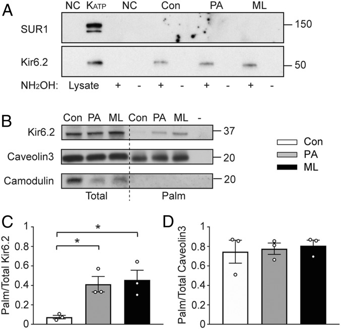
A physiological role for long-chain acyl-CoA esters to activate ATP-sensitive K+ (KATP) channels is well established. Circulating palmitate is transported into cells and converted to palmitoyl-CoA, which is a substrate for palmitoylation. We found that palmitoyl-CoA, but not palmitic acid, activated the channel when applied acutely. We have altered the palmitoylation state by preincubating cells with micromolar concentrations of palmitic acid or by inhibiting protein thioesterases. With acyl-biotin exchange assays we found that Kir6.2, but not sulfonylurea receptor (SUR)1 or SUR2, was palmitoylated. These interventions increased the KATP channel mean patch current, increased the open time, and decreased the apparent sensitivity to ATP without affecting surface expression. Similar data were obtained in transfected cells, rat insulin-secreting INS-1 cells, and isolated cardiac myocytes. Kir6.2ΔC36, expressed without SUR, was also positively regulated by palmitoylation. Mutagenesis of Kir6.2 Cys166 prevented these effects. Clinical variants in KCNJ11 that affect Cys166 had a similar gain-of-function phenotype, but was more pronounced. Molecular modeling studies suggested that palmitoyl-C166 and selected large hydrophobic mutations make direct hydrophobic contact with Kir6.2-bound PIP2 Patch-clamp studies confirmed that palmitoylation of Kir6.2 at Cys166 enhanced the PIP2 sensitivity of the channel. Physiological relevance is suggested since palmitoylation blunted the regulation of KATP channels by α1-adrenoreceptor stimulation. The Cys166 residue is conserved in some other Kir family members (Kir6.1 and Kir3, but not Kir2), which are also subject to regulated palmitoylation, suggesting a general mechanism to control the open state of certain Kir channels.

Keywords: KATP channels; Kir6.2; PIP2; lipidation; palmitoylation.


Conflict of interest statement

The authors declare no competing interest.

Figures

Fig. 1.
Fig. 1.
KATP channels are palmitoylation targets. (A) The Kir6.2 but not SUR1 subunit is palmitoylated. Lysates of HEK-293 cells cotransfected with Kir6.2-myc/Flag-SUR1 were subjected to acyl-biotin exchange assays. Negative control reactions included untransfected cells (NC) or reactions without hydroxylamine (NH2OH), in which the Cys-palmitoyl thioester linkages remain intact and cannot bind to biotin. Representative immunoblot of biotinylated proteins to detect SUR1 (with an anti-Flag antibody) or Kir6.2 (with an anti-myc antibody). The left two lanes are cell lystes. (B) Acyl-biotin exchange assay showed Kir6.2 subunit is palmitoylated in cultured rat adult cardiomyocytes, where “-” indicates a Tris-treated group as the negative experimental control and hydroxylamine was applied to control, PA, and ML groups. Caveolin3 and camodulin were used as positive and negative control, respectively. (C) Summary data of the stoichiometry of palmitoylated Kir6.2 to total Kir6.2. (D) Summary data of the stoichiometry of palmitoylated to total caveolin3. n = 3 blots per group. *P < 0.05 vs. control determined by Dunnett’s test following the ANOVA with repeated measures. Note that wild-type Kir6.2 migrates at ∼37 kDa (e.g., in B), whereas Kir6.2-myc, which has six C-terminal myc epitopes, migrates at a ∼50 kDa (e.g., in A).
Fig. 2.
Fig. 2.
Palmitoylation modulates endogenous KATP channels. (A) Representative inside-out current recordings obtained from cardiac myocytes in control, PA, and ML groups. ATP concentrations were switched as indicated. The mean patch current was recorded at a membrane potential of −80 mV and defined by the current component blocked by 1 mM ATP applied to the cytosolic face of the patch. The ATP-sensitivity of KATP channels was determined by plotting the KATP current (normalized to the maximum current) as a function of the cytosolic ATP concentration. Data from individual patches were subjected to curve fitting to a modified Boltzmann equation, yielding IC50 values for ATP inhibition. (B) Summary data of mean patch currents and IC50 of ATP-sensitivity for control, PA, and ML groups in cardiomyocytes. n ≥ 6 patches in each group. *P < 0.05, **P < 0.01 vs. the control group determined by Dunnett’s test following the ANOVA with repeated measures.
Fig. 3.
Fig. 3.
Palmitoylation of Kir6.2 at position Cys166 regulates KATP channel function. (A) Side view of Kir6.2 tetramer, with one Cys166 highlighted in red (PDB ID code 6C3P). (B) Representative immunoblots of acyl-biotin exchange assay and (C) The normalized ratios of palmitoylated Kir6.2 to total Kir6.2 are shown for wild-type and C166V mutated KATP channels transfected in HEK-293 cells. n = 3 blots per group. *P < 0.05 vs. control determined by Dunnett’s test following the ANOVA with repeated measures. Summary data of mean patch currents for Kir6.2-ΔC36 (D) and Kir6.2/SUR1 (E) channels carrying C166V mutation. n ≥ 11 patches in each group.
Fig. 4.
Fig. 4.
Palmitoylation enhances KATP channel PIP2 sensitivity. (A) Representative inside-out current recordings obtained from cardiomyocytes in control, PA, and ML groups. PIP2–diC8 concentrations were switched as indicated right after Ca2+ induced rundown. (B) Fractional currents (recovered currents normalized by the baseline current) for control, PA, and ML groups in cardiomyocytes was plotted as a function of PIP2–diC8 concentration and subjected to curve fitting to a modified Hill equation. Summary data of fractional currents for 1 µM (C) and 10 µM (D) PIP2–diC8 in HEK-293 cells transfected with either wild-type or C166V mutated KATP channels. n ≥ 6 patches in each group. *P < 0.05, **P < 0.01 vs. control determined by Dunnett’s test following the ANOVA with repeated measures.
Fig. 5.
Fig. 5.
Palmitoylation desensitizes KATP channel to membrane PIP2 change. (A) Representative whole-cell KATP current recordings from PA treated HEK-293 cells transfected with α1-adrenoreceptor and either wild-type or C166V mutated KATP channels (Kir6.2/SUR2A). Pinacidil and MTX are applied as indicated. Percentage of remaining (minimum current recorded at the presence of MTX normalized by the maximum current with pinacidil only) for control, PA, and ML groups in cardiomyocytes (B) and HEK-293 cells transfected with either wild-type or C166V mutated KATP channels (C). n ≥ 3 patches in each group. *P < 0.05, **P < 0.01 vs. control determined by Dunnett’s test following the ANOVA with repeated measures.
Fig. 6.
Fig. 6.
KCNJ11 variants at Cys166 associated with genetic disorders also regulate KATP channel activity. (A) Summary data of mean patch currents for wild-type KATP channel and channels carrying C166F and C166Y mutations. (B) Fractional currents (normalized by the mean patch current in the absence of ATP) were plotted as a function of ATP concentration and subjected to curve fitting to a modified Hill equation for wild-type KATP channel and channels carrying C166F and C166Y mutations. n ≥ 3 patches in each group. *P < 0.05, **P < 0.01, ***P < 0.005 vs. wild-type determined by Dunnett’s test following the ANOVA with repeated measures. (C) Representative inside-out current recordings from wild-type and C166Y KATP channels, perfused with 100 μM Ca2+, 10 μM PIP2-diC8 and 1 mM Ba2+. Dashed lines indicate closed state. (D) Summarized data of percentage of Ca2+-induced rundown for wild-type and C166Y KATP channels. n ≥ 4 patches in each group. ***P < 0.005 vs. wild-type determined by unpaired t test.
Fig. 7.
Fig. 7.
C166-bound palmitate chain can directly contact PIP2. (A) Side view of the Kir6.2 pore-forming tetramer (each monomer is displayed in ribbon depiction in a different color: Purple, yellow, green and gray) in the open conformation (PDB ID code 3SYQ) with Cys166 from the green monomer palmitoylated and the PIP2 binding site from the purple monomer displayed. The carbon chain of palmitoyl-C166 contacts the hydrophobic ether moeities of PIP2. A model of the green monomer based on the closed state (PDB ID code 63CP) is superimposed. (B) Magnified same view as in A of the palmitoyl-C166 interaction with PIP2, showing the expected projections of C166F, Y, and V. (C) View perpendicular to the membrane from the intracellular face of the pore of Kir6.2 showing the proximity of the inner helix gate to the palmitoyl-C166 interaction with PIP2 at the cytosolic membrane interface.
Fig. 8.
Fig. 8.
Conserved palmitoylation site across Kir channels. (A) Of the human inward rectifier K+ channel (Kir) subunits, only the Kir3.x and Kir6.x subfamilies have a conserved cysteine residue at the inner membrane interface (dark box). Shown are sequence alignments of Kirx.x subunits of selected regions that span the inner membrane interface. The light blue boxes, respectively, indicate the start and end of the M1 and M2 transmembrane helixes. Shading is by group conservation. The boxed Cys is the putative palmitoylation site. (B) Representative acyl-biotin exchange assay immunoblots and the ratios of palmitoylated to total Kir subunits for Kir6.1, Kir3.4, Kir2.1, and Kir2.1-A178C. n ≥ 3 blots per group. *P < 0.05 vs. control determined by unpaired t test.

References

    1. Lin D. T., Conibear E., Enzymatic protein depalmitoylation by acyl protein thioesterases. Biochem. Soc. Trans. 43, 193–198 (2015). - PubMed
    1. Lin D. T., Conibear E., ABHD17 proteins are novel protein depalmitoylases that regulate N-Ras palmitate turnover and subcellular localization. eLife 4, e11306 (2015). - PMC - PubMed
    1. Cox A. D., Der C. J., Philips M. R., Targeting RAS membrane association: Back to the future for anti-RAS drug discovery? Clin. Cancer Res. 21, 1819–1827 (2015). - PMC - PubMed
    1. Aicart-Ramos C., Valero R. A., Rodriguez-Crespo I., Protein palmitoylation and subcellular trafficking. Biochim. Biophys. Acta 1808, 2981–2994 (2011). - PubMed
    1. Tortosa E., Hoogenraad C. C., Polarized trafficking: The palmitoylation cycle distributes cytoplasmic proteins to distinct neuronal compartments. Curr. Opin. Cell Biol. 50, 64–71 (2018). - PubMed

Publication types

MeSH terms

LinkOut - more resources