. 2020 Apr 24;10(1):6963.
doi: 10.1038/s41598-020-63922-8.

Choosing the Right Differentiation Medium to Develop Mucociliary Phenotype of Primary Nasal Epithelial Cells In Vitro

Affiliations

Choosing the Right Differentiation Medium to Develop Mucociliary Phenotype of Primary Nasal Epithelial Cells In Vitro

Anja E Luengen et al. Sci Rep. .

Abstract

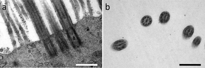
In vitro differentiation of airway epithelium is of interest for respiratory tissue engineering and studying airway diseases. Both applications benefit from the use of primary cells to maintain a mucociliated phenotype and thus physiological functionality. Complex differentiation procedures often lack standardization and reproducibility. To alleviate these shortfalls, we compared differentiation behavior of human nasal epithelial cells in four differentiation media. Cells were differentiated at the air-liquid interface (ALI) on collagen-coated inserts. Mucociliary differentiation status after five weeks was analyzed by electron microscopy, histology and immunohistochemistry. The amount of ciliation was estimated and growth factor concentrations were evaluated using ELISA. We found that retinoic-acid-supplemented mixture of DMEM and Airway Epithelial Cell Growth Medium gave most promising results to obtain ciliated and mucus producing nasal epithelium in vitro. We discovered the balance between retinoic acid (RA), vascular endothelial growth factor (VEGF), epidermal growth factor (EGF) and fibroblast growth factor β (FGF-β) to be relevant for differentiation. We could show that low VEGF, EGF and FGF-β concentrations in medium correspond to absent ciliation in specific donors. Therefore, our results may in future facilitate donor selection and non-invasive monitoring of ALI cultures and by this contribute to improved standardization of epithelial in vitro culture.


Conflict of interest statement

The authors declare no competing interests.

Figures

Figure 1
Figure 1
Ciliation after four weeks of ALI in different media (SEM). (ah) SEM revealed cilia formation using mAir and microvilli formation without ciliation in Pneu-, Epi- and EMM-cultures. Representative images of two different magnifications are shown. Scale bar: 5 µm.
Figure 2
Figure 2
Cilia analysis of mAir-cultures (TEM). (a) Detection of basal bodies confirmed cilia formation; (b) Identification of central microtubuli doublets verified presence of secondary cilia. Representative images are shown. Scale bar: 500 nm.
Figure 3
Figure 3
Ciliation scoring in ALI-cultures using different media. Scoring results showed statistically significant differences between all media. Results are expressed as mean ± SD following Gaussian error propagation. Statistically significant differences (p < 0.05) are indicated by *.
Figure 4
Figure 4
PAS reaction of mucociliary differentiation. (a,f) PAS reaction confirmed presence of glycogens, mucopolysaccharides and glycoproteins in human nasal concha tissue; (be,gj) PAS reaction indicated cell multilayers and confirmed mucus production in mAir-cultures while using other media led to cell monolayers lacking mucociliary differentiation. Representative pictures of two different magnifications are shown. Scale bar: 40 µm.
Figure 5
Figure 5
Immunohistochemical staining for differentiation status after four weeks of ALI in different media. (ae,k) Claudin-1 (green) indicated tight junction formation in human nasal concha tissue and ALI-cultures. α-tubulin (red) visualized cilia in native tissue and mAir-cultures; (fj,l) pan-cytokeratin (green) detected a wide range of keratins in all cultures and Mucin5AC (red) showed presence of goblet cells and mucus in the native control and in cell layers cultured in mAir. DAPI (blue) stained cell nuclei. Representative pictures are shown. Scale bar: 20 µm.
Figure 6
Figure 6
ELISA for human growth factor and RA analysis. (a) Concentrations of growth factors VEGF, EGF, PDGF-BB, β-NGF, SCF, TNF-α, FGF-β and TGF-β in different media in ALI after four weeks (selected donors); (b) VEGF, EGF and FGF-β concentrations from (a) normalized to cell-free medium (selected donors); (c) RA concentrations in different media in ALI after four weeks (all donors); (d) RA concentrations normalized to cell-free medium (all donors); The absorbance was measured at a wavelength of 450 nm. Error bars show standard deviation. Statistical significance indicated by * shows significance (p < 0.05) between mean values.
Figure 7
Figure 7
Comparison of growth factor and RA concentrations between ciliated and non-ciliated donors. (a) Growth factor concentrations for VEGF, EGF and FGF-β in mAir and EMM; (b) RA concentrations in mAir and EMM; Unfilled symbols and dashed bars represent donors without ciliation in any medium. Error bars show standard deviation. Statistical significance indicated by * shows significance (p < 0.05) between mean values.

References

    1. Tilley AE, Walters MS, Shaykhiev R, Crystal RG. Cilia dysfunction in lung disease. Annual review of physiology. 2015;77:379–406. doi: 10.1146/annurev-physiol-021014-071931. - DOI - PMC - PubMed
    1. Matthys H, Vastag E, Köhler D, Daikeler G, Fischer J. Mucociliary clearance in patients with chronic bronchitis and bronchial carcinoma. Respiration. 1983;44:329–337. doi: 10.1159/000194565. - DOI - PubMed
    1. Folch E, Keyes C. Airway stents. Annals of cardiothoracic surgery. 2018;7:273. doi: 10.21037/acs.2018.03.08. - DOI - PMC - PubMed
    1. Chin CS, Litle V, Yun J, Weiser T, Swanson SJ. Airway stents. Ann Thorac Surg. 2008;85:S792–6. doi: 10.1016/j.athoracsur.2007.11.051. - DOI - PubMed
    1. Etienne, H. et al. Tracheal replacement. European Respiratory Journal51 (2018). - PubMed

Publication types

MeSH terms