. 2020 Apr 24;10(1):6959.
doi: 10.1038/s41598-020-63886-9.

Bridging repair of the abdominal wall in a rat experimental model. Comparison between uncoated and polyethylene oxide-coated equine pericardium meshes

Affiliations

Bridging repair of the abdominal wall in a rat experimental model. Comparison between uncoated and polyethylene oxide-coated equine pericardium meshes

Alessandro Pasculli et al. Sci Rep. .

Abstract

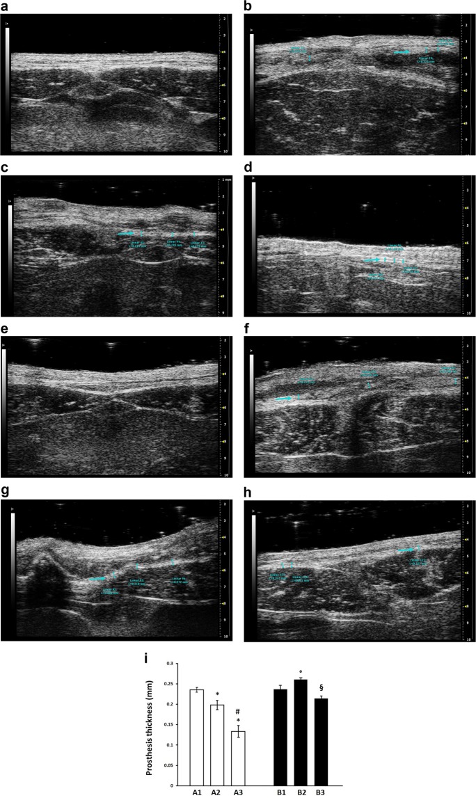
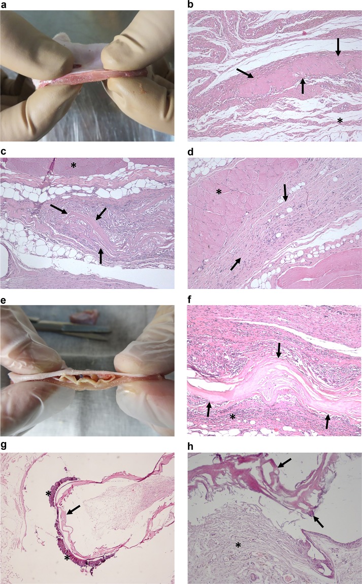
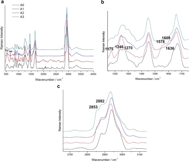
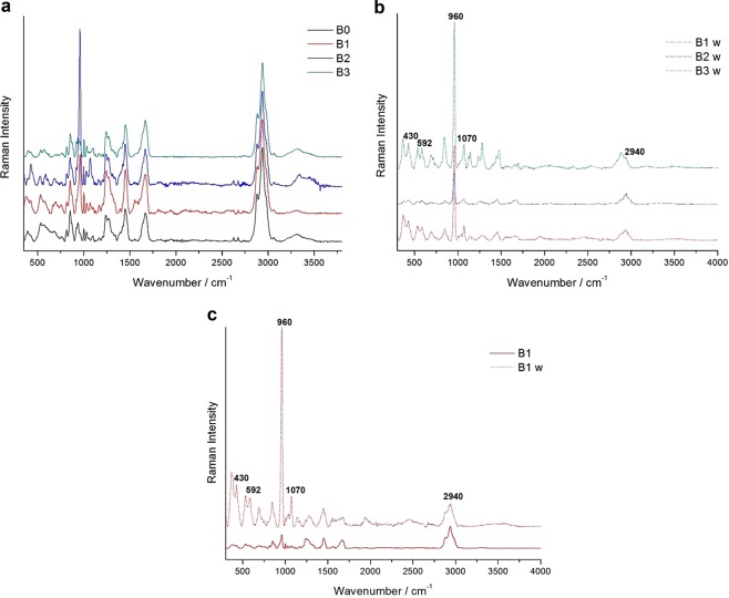
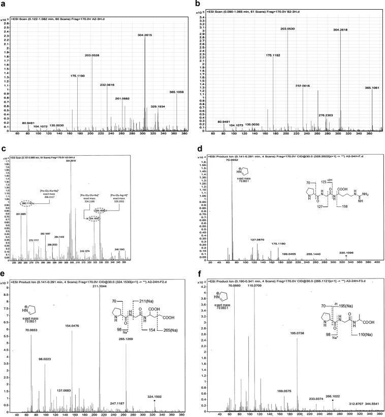
Biological meshes improve the outcome of incisional hernia repairs in infected fields but often lead to recurrence after bridging techniques. Sixty male Wistar rats undergoing the excision of an abdominal wall portion and bridging mesh repair were randomised in two groups: Group A (N = 30) using the uncoated equine pericardium mesh; Group B (N = 30) using the polyethylene oxide (PEO)-coated one. No deaths were observed during treatment. Shrinkage was significantly less common in A than in B (3% vs 53%, P < 0.001). Adhesions were the most common complication and resulted significantly higher after 90 days in B than in A (90% vs 30%, P < 0.01). Microscopic examination revealed significantly (P < 0.05) higher mesh integrity, fibrosis and calcification in B compared to A. The enzymatic degradation, as assessed with Raman spectroscopy and enzyme stability test, affected A more than B. The PEO-coated equine pericardium mesh showed higher resistance to biodegradation compared to the uncoated one. Understanding the changes of these prostheses in a surgical setting may help to optimize the PEO-coating in designing new biomaterials for the bridging repair of the abdominal wall.


Conflict of interest statement

The authors declare no competing interests.

Figures

Figure 1
Figure 1
(ad) Ultrasonography sample images of B-mode acquisition of a Group A rat abdomen at the level of the surgical scar. (a) before the implant. (b) 30 days after the implant (A1). (c) 60 days after the implant (A2). (d) 90 days after the implant (A3). The blue arrows identify the mesh. (eh) Ultrasonography sample image of B-mode acquisition of the Group B rat abdomen at the level of the surgical scar. (e) before the implant. (f) 30 days after the implant (B1). (g) 60 days after the implant (B2). (h) 90 days after the implant (B3). The blue arrows identify the mesh. (i) Prosthesis thickness measured by ultrasound in each experimental group. Each bar is the mean ± standard deviation from five to nine measurements. Statistical analysis by unpaired Student’s t-test indicates P < 0.05 versus A1 group (*), P < 0.01 versus A2 group (#), P < 0.05 versus B1 group (°) and P = 0.001 versus B2 group (§).
Figure 2
Figure 2
(ad) Group A: (a) Group A1 prosthesis soon after its removal from the rat. (b) Group A1 10x hematoxylin-eosin stained section, the arrows show a fragment of the prosthesis, the asterisk identifies subcutaneous tissue. (c) Group A2 10x hematoxylin-eosin stained section, the arrows indicate a fragment of the prosthesis, the asterisk identifies the muscular layer. (d) Group A3 10x hematoxylin-eosin stained section, the prosthesis is no longer recognisable and is substituted by fibrous tissue (arrows), the asterisk identifies the muscular layer. (eh) Group B: (e) Group B1 prosthesis soon after its removal from the rat. (f) Group B1 10x hematoxylin-eosin stained section, intact prosthesis (arrows) encircled by inflammatory infiltrate (asterisk). (g) Group B2 4x hematoxylin-eosin stained section, calcification (asterisks) on the edge of the prosthesis (arrow). (h) Group B3 10x hematoxylin-eosin stained section, the prosthesis is still visible (arrow) and appears to be encircled by fibrous tissue (asterisk).
Figure 3
Figure 3
Representative baseline-subtracted Raman spectra of Group A prostheses before and after surgery in the 500–4000 cm−1 region (a) and details of the 1100–1800 (b) and 2600–3200 cm−1 (c) regions. The intensities are normalised to the band at 2940 cm−1, the spectra are shifted on the Y-axis for better readability. The peaks cited in the text are marked.
Figure 4
Figure 4
Representative baseline-subtracted Raman spectra of Group B prostheses before and after surgery (a), after surgery on the white side (b), and comparison between B1 on the two sides (c). For each single group of spectra, the intensities are normalised to the band at 2940 cm−1, the spectra are shifted on the Y-axis for better readability. The peaks cited in the text are marked.
Figure 5
Figure 5
HRMS and tandem MS/MS fragmentation traces of representative supernatant samples of prostheses at different times of incubation in digest solutions at 37 °C: (a) MS spectrum up to 390 m/z (ESI scan recorded up to 1200 m/z, positive mode) of a representative Group A prosthesis sample after 3 h incubation; (b) MS spectrum up to 390 m/z (ESI scan recorded up to 1200 m/z, positive mode) of a representative Group B prosthesis after 3 h incubation; (c) putative structure assessment of some m/z signals of a representative Group A sample after 24 h incubation; tentative assignments by MS/MS fragmentation analysis of the signals at (d) m/z 329 (Pro-Gly-Arg), (e) m/z 324 signal (Pro-Gly-Glu) and (f) m/z 266 signal (Pro-Gly-Ala).

References

    1. Mudge M, Hughes LE. Incisional hernia: a 10 year prospective study of incidence and attitudes. Br. J. Surg. 1985;72:70–71. doi: 10.1002/bjs.1800720127. - DOI - PubMed
    1. Penttinen R, Grönroos JM. Mesh repair of common abdominal hernias: a review on experimental and clinical studies. Hernia. 2008;12:337–344. doi: 10.1007/s10029-008-0362-4. - DOI - PubMed
    1. Choi JJ, et al. Use of mesh during ventral hernia repair in clean-contaminated and contaminated cases: outcomes of 33,832 cases. Ann. Surg. 2012;255:176–180. doi: 10.1097/SLA.0b013e31822518e6. - DOI - PubMed
    1. Usher FC, Ochsner J, Tuttle LL., Jr. Use of marlex mesh in the repair of incisional hernias. Am. Surg. 1985;24:969–974. - PubMed
    1. den Hartog D, Dur AH, Tuinebreijer WE, Kreis RW. Open surgical procedures for incisional hernias. Cochrane Database Syst. Rev. 2008;3:CD006438. - PMC - PubMed

Substances