. 2021 Jan;78(2):675-693.
doi: 10.1007/s00018-020-03514-x. Epub 2020 Apr 24.

Fucosidases from the human gut symbiont Ruminococcus gnavus

Affiliations

Fucosidases from the human gut symbiont Ruminococcus gnavus

Haiyang Wu et al. Cell Mol Life Sci. 2021 Jan.

Abstract

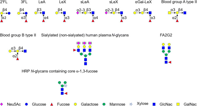
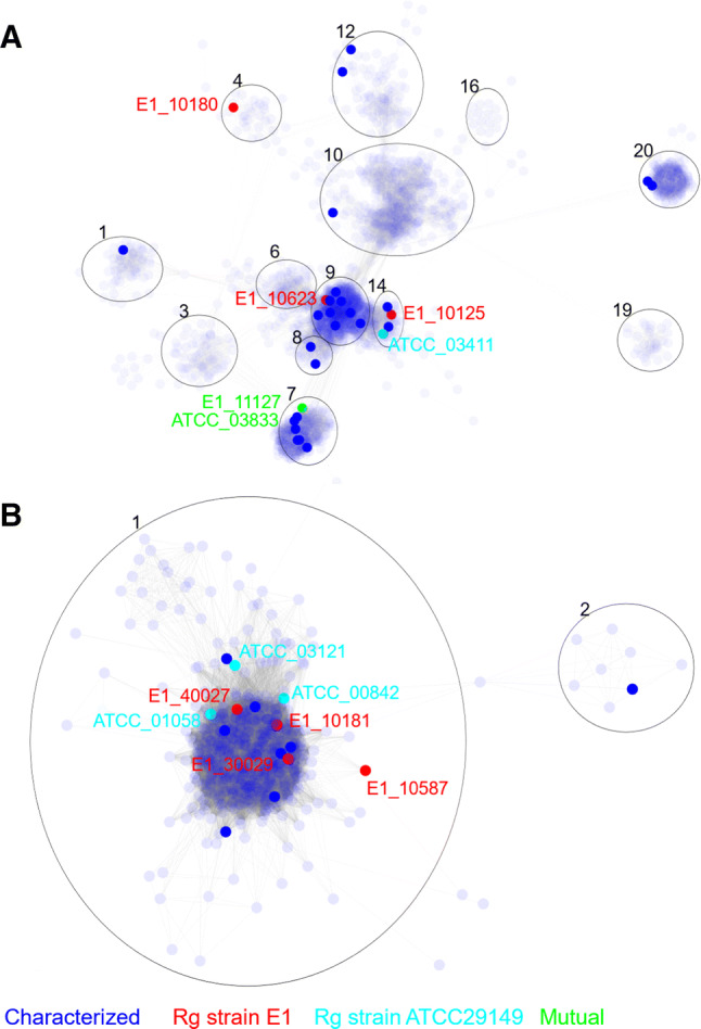
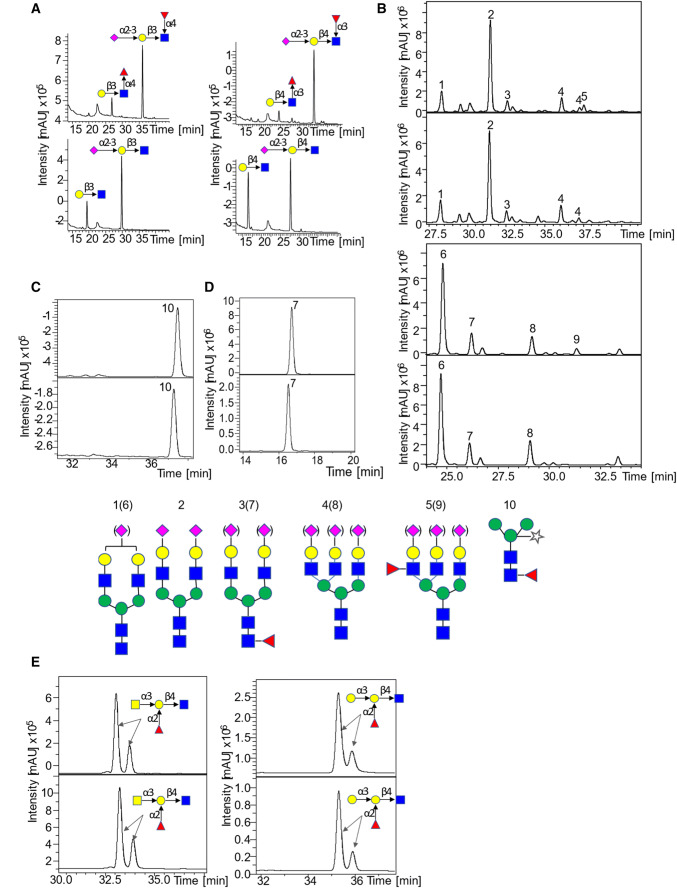
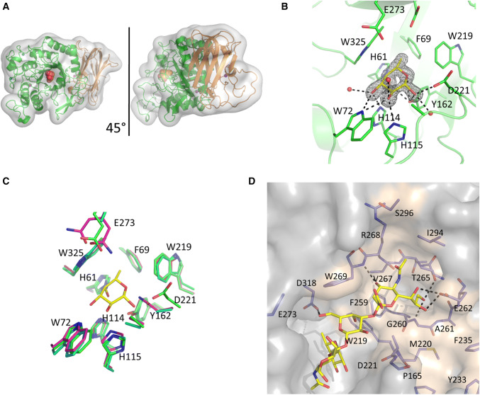
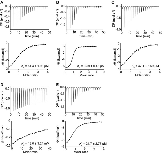
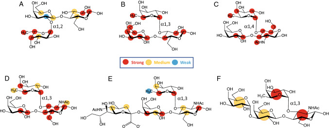
The availability and repartition of fucosylated glycans within the gastrointestinal tract contributes to the adaptation of gut bacteria species to ecological niches. To access this source of nutrients, gut bacteria encode α-L-fucosidases (fucosidases) which catalyze the hydrolysis of terminal α-L-fucosidic linkages. We determined the substrate and linkage specificities of fucosidases from the human gut symbiont Ruminococcus gnavus. Sequence similarity network identified strain-specific fucosidases in R. gnavus ATCC 29149 and E1 strains that were further validated enzymatically against a range of defined oligosaccharides and glycoconjugates. Using a combination of glycan microarrays, mass spectrometry, isothermal titration calorimetry, crystallographic and saturation transfer difference NMR approaches, we identified a fucosidase with the capacity to recognize sialic acid-terminated fucosylated glycans (sialyl Lewis X/A epitopes) and hydrolyze α1-3/4 fucosyl linkages in these substrates without the need to remove sialic acid. Molecular dynamics simulation and docking showed that 3'-Sialyl Lewis X (sLeX) could be accommodated within the binding site of the enzyme. This specificity may contribute to the adaptation of R. gnavus strains to the infant and adult gut and has potential applications in diagnostic glycomic assays for diabetes and certain cancers.

Keywords: Antennary fucose; Glycoside hydrolase; Gut microbiota; Lewis epitopes; Mucin glycosylation; Mucus.


Figures

Fig. 1
Fig. 1
Fucosylated oligosaccharides used in this study. Monosaccharide symbols follow the Symbol Nomenclature for Glycans system [98]
Fig. 2
Fig. 2
The distribution of R. gnavus GH29 and GH95 fucosidases based on SSN analysis. a Partial representation of SSN analysis of GH29 family containing fucosidases from R. gnavus E1 and ATCC29149 strains. b Representation of the SSN central cluster of GH95 family containing all GH95 from R. gnavus E1 and ATCC29149 strains. Blue node: sequences extracted from the CAZy database encoding functionally characterized enzymes. Red nodes sequences from R. gnavus E1 strain. Cyan nodes, sequences from R. gnavus ATCC29149 strain. Green nodes, sequences common to both R. gnavus E1 and ATCC29149 strains
Fig. 3
Fig. 3
LC–MS/MS analysis of R. gnavus GH29 fucosidase E1-10125 towards various fucosylated substrates. a LC–MS/MS analysis of the products released from the enzymatic reaction of E1-10125 with LeA (left) and LeX (right), the upper graph is the negative control and the lower one corresponds to the enzymatic reaction. B LC–MS analysis of the products released from the enzymatic reaction of E1-10125 with sialylated (upper) and desialylated (lower) human plasma. The negative controls are showed on top of the enzymatic reactions. c LC–MS analysis of the products released from the enzymatic reaction of E1-10125 with HRP (core α1,3-fucose) (lower). The negative control is shown in the upper panel. D. LC–MS analysis of the products released from the enzymatic reaction of E1-10125 with FA2G2 (α1,6-fucose) (lower). The negative control is shown in the upper panel. e LC–MS analysis of the products released from the enzymatic reaction of E1-10125 with blood group A type II (left) and blood group B type II (right) (upper). The negative control is shown in the upper panel
Fig. 4
Fig. 4
Crystal structure of R. gnavus GH29 fucosidase E1_10125. a Cartoon representation of E1_10125 fucosidase, the catalytic domain is coloured green and the proposed CBM is coloured orange. A fucose residue in a sphere representation indicates the location of the active site. The views are related by a 45° rotation around the y axis. b The E1_10125 fucose binding site. The β-anomer of fucose is shown in yellow with nearby active site residues shown in green. Black dashed lines indicate hydrogen-bonding interactions. Fo–Fc difference map density for the fucose residue is displayed as a black mesh, contoured at 2σ. c The fucose binding sites of E1_10125 (green), S. pneumoniae GH29 fucosidase (magenta), and B. longum subsp. infantis GH29 fucosidase (cyan) are aligned. Residue numbers refer to E1_10125. The binding site residues are conserved across the three structures and differences present at the D221 and E273 positions are catalytic mutants. Fucose bound in the E1_10125 is show in yellow for reference. d Model of the orientation and conformation of sLeX bound to R. gnavus E1_10125 proposed by MD simulations
Fig. 5
Fig. 5
ITC isotherms of R. gnavus GH29 fucosidase E1_10125 binding to fucosylated ligands. a E1_10125 binding to LeX. b E1_10125 binding to sLeX. c E1_10125 binding to αGal-LeX. d E1_10125 binding to Neu5Ac. e E1_10125 binding to l-Fucose. DP differential power
Fig. 6
Fig. 6
Binding epitope mapping from STD NMR spectroscopy depicting interactions of R. gnavus GH29 fucosidase E1-10125 with fucosylated oligosaccharides. Normalized saturation transfer intensities (0–100%) from STD NMR experiments mapped onto the chemical structures of a 2′FL, b 3FL, c LeA, d LeX, e sLeX and f αGal-LeX. Stronger normalized STD intensities correlate with closer ligand contacts with the surface of the protein in the bound state. Legend indicates normalized STD intensities: blue, 0–24%; yellow, 25–50%, red 51–100%. The enzyme intimately recognizes 3FL and LeA, whereas looser contacts are observed for 2′FL, LeX, sLeX and αGal-LeX. For the latter, a much higher degree of proton chemical shift overlapping implied lower binding epitope resolution and a normalized STD intensity value was assigned for each ring, as an average of the STD intensities of its isolated protons

References

    1. Thursby E, Juge N. Introduction to the human gut microbiota. Biochem J. 2017;474(11):1823–1836. doi: 10.1042/BCJ20160510. - DOI - PMC - PubMed
    1. Bode L. Human milk oligosaccharides: every baby needs a sugar mama. Glycobiology. 2012;22(9):1147–1162. doi: 10.1093/glycob/cws074. - DOI - PMC - PubMed
    1. Cabrera-Rubio R, Kunz C, Rudloff S, Garcia-Mantrana I, Crehua-Gaudiza E, Martinez-Costa C, Collado MC. Association of maternal secretor status and human milk oligosaccharides with milk microbiota: an observational pilot study. J Pediatr Gastr Nutr. 2019;68(2):256–263. doi: 10.1097/Mpg.0000000000002216. - DOI - PubMed
    1. Koropatkin NM, Cameron EA, Martens EC. How glycan metabolism shapes the human gut microbiota. Nat Rev Microbiol. 2012;10(5):323–335. doi: 10.1038/nrmicro2746. - DOI - PMC - PubMed
    1. Tailford LE, Crost EH, Kavanaugh D, Juge N. Mucin glycan foraging in the human gut microbiome. Front Genet. 2015;6:ARTN81. doi: 10.3389/fgene.2015.00081. - DOI - PMC - PubMed

MeSH terms

Supplementary concepts

LinkOut - more resources