

Contrast enhanced ultrasonography (CEUS) to detect abdominal microcirculatory disorders in severe cases of COVID-19 infection: First experience
- PMID: 32333581
- PMCID: PMC7369109
- DOI: 10.3233/CH-209003
Contrast enhanced ultrasonography (CEUS) to detect abdominal microcirculatory disorders in severe cases of COVID-19 infection: First experience
Abstract
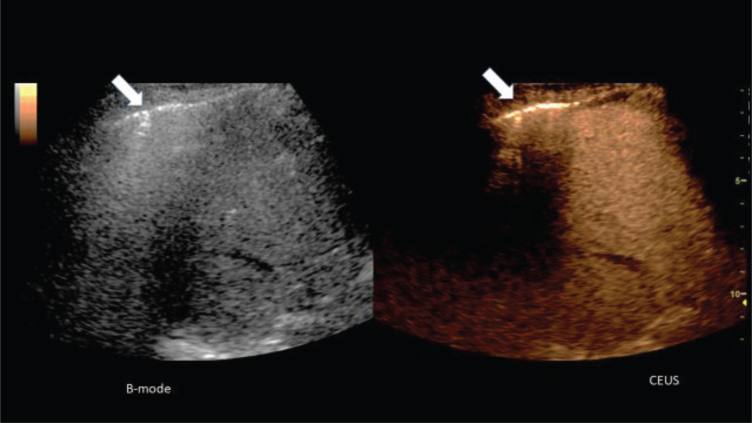
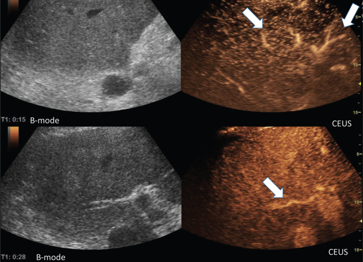
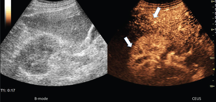
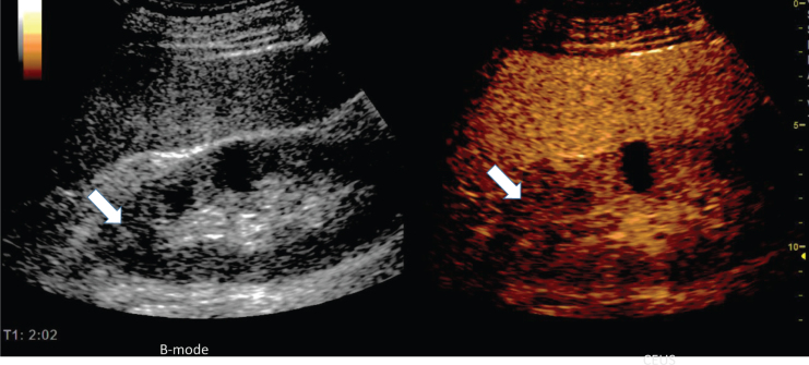
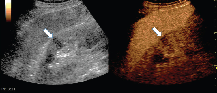
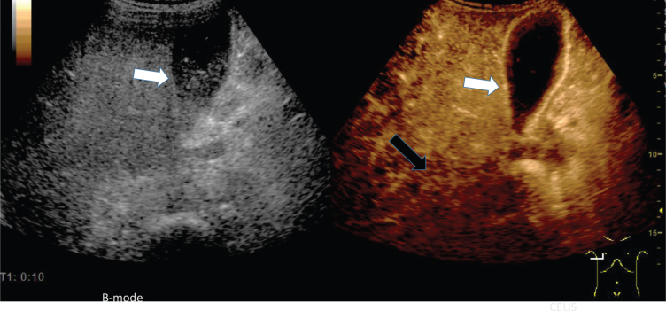
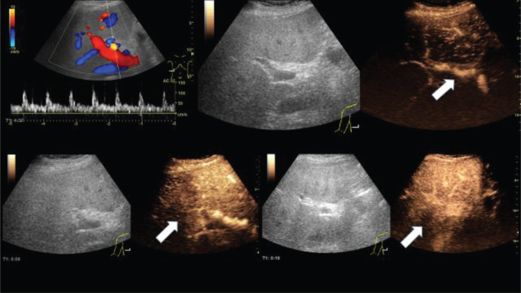
In the hands of experienced examiners, the contrast enhanced sonography (CEUS) offers the possibility to analyze dynamic microcirculatory disturbances in real time dynamically without any risk for kidneys and thyroid gland even in severe progressing disease bedside. Based on severe COVID-19 infections, first experiences with abdominal CEUS examinations are presented. In the stage of an imminent organ failure with significantly reduced kidney and liver function, CEUS can be used to show a narrowing of the organ-supplying arteries, as well as a delayed capillary filling of vessels near the capsule, a regional reduced parenchymal perfusion or an inflammatory hyperemia with capillary hypercirculation. It is possible to quickly rule out organ infarction and to dynamically record the mesenteric arterial and venous blood flow.
Keywords: COVID-19; Corona virus; contrast enhanced ultrasound sonography (CEUS); kidney; liver.
Figures
Comment in
-
Complement-mediated Extracellular Vesicle release as a measure of endothelial dysfunction and prognostic marker for COVID-19 in peripheral blood - Letter to the Editor.Clin Hemorheol Microcirc. 2020;75(4):383-386. doi: 10.3233/CH-200958. Clin Hemorheol Microcirc. 2020. PMID: 32925002 No abstract available.
References
-
- Zhang T, Sun LX, Feng RE. Comparison of Clinical and Pathological Features Between Severe Acute Respiratory Syndrome and Coronavirus Disease 2019. Zhonghua Jie He He Hu Xi Za Zhi. 2020;43:E040. - PubMed
-
- Yang X, Yu Y, Xu J, Shu H. Jia’an. Clinical course and outcomes of critically ill patients with SARS-CoV-2 pneumonia in Wuhan, China: a single-centered, retrospective, observational study. Lancet. 2020. doi.org/10.1016/S2213-2600(20)30079-5 - DOI - PMC - PubMed
-
- Han R, Huang L, Jiang H, Dong J, Peng H, Zhang D. Early Clinical and CT Manifestations of Coronavirus Disease 2019 (COVID-19) Pneumonia. AJR. 2020;215:1–6. - PubMed
-
- Zhang HW, Yu J, Xu HJ, Lei Y, Pu ZH, Dai WC, Lin F, Wang YL, Wu XL, Liu LH, Li M, Mo YQ, Zhang H, Luo SP, Chen H, Lyu GW, Zhou ZG, Liu WM, Liu XL, Song HY, Chen FZ, Zeng L, Zhong H, Guo TT, Hu YQ, Yang XX, Liu PN, Li DF. Corona Virus International Public Health Emergencies: Implications for Radiology Management. Acad Radiol. 2020;27(4):463–7. - PMC - PubMed
MeSH terms
Substances
LinkOut - more resources
Full Text Sources
Other Literature Sources
Medical
