. 2020 May 12;26(6):797-808.
doi: 10.1093/ibd/izaa085.

Expression of SARS-CoV-2 Entry Molecules ACE2 and TMPRSS2 in the Gut of Patients With IBD

Affiliations

Expression of SARS-CoV-2 Entry Molecules ACE2 and TMPRSS2 in the Gut of Patients With IBD

Juan F Burgueño et al. Inflamm Bowel Dis. .

Abstract

Background: Patients with inflammatory bowel disease (IBD) have intestinal inflammation and are treated with immune-modulating medications. In the face of the coronavirus disease-19 pandemic, we do not know whether patients with IBD will be more susceptible to infection or disease. We hypothesized that the viral entry molecules angiotensin I converting enzyme 2 (ACE2) and transmembrane serine protease 2 (TMPRSS2) are expressed in the intestine. We further hypothesized that their expression could be affected by inflammation or medication usage.

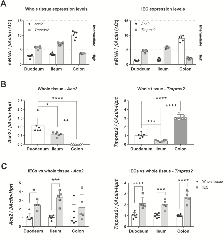
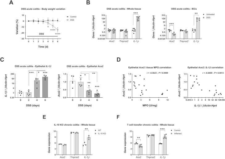
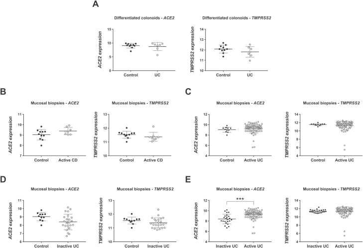
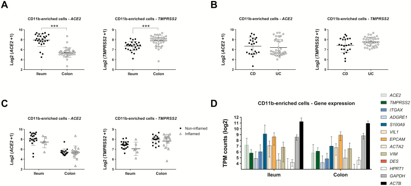
Methods: We examined the expression of Ace2 and Tmprss2 by quantitative polymerase chain reacion in animal models of IBD. Publicly available data from organoids and mucosal biopsies from patients with IBD were examined for expression of ACE2 and TMPRSS2. We conducted RNA sequencing for CD11b-enriched cells and peripheral and lamina propria T-cells from well-annotated patient samples.

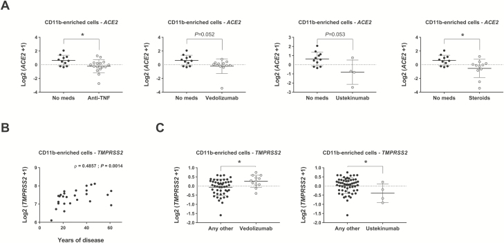
Results: ACE2 and TMPRSS2 were abundantly expressed in the ileum and colon and had high expression in intestinal epithelial cells. In animal models, inflammation led to downregulation of epithelial Ace2. Expression of ACE2 and TMPRSS2 was not increased in samples from patients with compared with those of control patients. In CD11b-enriched cells but not T-cells, the level of expression of ACE2 and TMPRSS2 in the mucosa was comparable to other functional mucosal genes and was not affected by inflammation. Anti-tumor necrosis factor drugs, vedolizumab, ustekinumab, and steroids were linked to significantly lower expression of ACE2 in CD11b-enriched cells.

Conclusions: The viral entry molecules ACE2 and TMPRSS2 are expressed in the ileum and colon. Patients with IBD do not have higher expression during inflammation; medical therapy is associated with lower levels of ACE2. These data provide reassurance for patients with IBD.

Keywords: Crohn disease; coronavirus; ulcerative colitis.


Figures

FIGURE 1.
FIGURE 1.
Ace2 and Tmprss2 expression in mouse gut and IECs in steady-state conditions. mRNA expression of Ace2 and Tmprss2 was analyzed by qPCR in different segments of the gut and in IECs isolated from these segments. A, Whole tissue expression levels of both genes in whole tissue or IECs as related to normal βActin expression. Note that lower ΔCt values mean higher expression levels, closer to those of βActin. N = 6 mice. B, Ace2 and Tmprss2 expression in whole tissue from selected gut locations. N = 6 mice; *P < 0.05, **P < 0.01, ***P < 0.001, and ****P < 0.0001 as determined by 1-way ANOVA followed by Tukey posthoc test. C, Ace2 and Tmprss2 in regional IECs normalized to their matched whole tissue expression. N = 5-6 mice; *P < 0.05, ***P < 0.001, and ****P < 0.0001 as determined by 2-way ANOVA followed by Sidak posthoc test. ANOVA indicates analysis of variance; qPCR, quantitative polymerase chain reaction.
FIGURE 2.
FIGURE 2.
Ace2 and Tmprss2 expression in mouse colon and IECs during inflammation. C57Bl/6 mice treated with 3% DSS for indicated times and expression of Ace2, Tmprss2, and IL-1β transcripts in the colon determined by qPCR. A, Body weight variation. N = 7-8 mice; *P < 0.05, ****P < 0.0001 as determined by 2-way ANOVA followed by Sidak posthoc test. B, Gene expression on day 6 of inflammation in whole colon and IECs isolated from colon. N = 7-8 (whole tissue) or n = 5 (colon IECs); **P < 0.01, ***P < 0.001 as determined by unpaired 2-tailed Student t test. C, Time course expression of IL-1β and Ace2 in colonic IECs isolated on days 0, 2, 4, and 6 of DSS treatment. N = 5 mice; IL-1β on day 6 vs day 0, ***P < 0.001; Ace2 on day 4 vs day 0, **P < 0.01; Ace2 on day 6 vs day 0, ***P < 0.001 as determined by 1-way ANOVA followed by Tukey posthoc test. D, Correlation between epithelial Ace2 expression levels and matched colon determinations of MPO (left panel) or epithelial IL-1β (right panel) on days 0, 2, 4, and 6 of DSS. N = 20 mice; Ace2/MPO, r = –0.5623, **P = 0.0099; Ace2/IL-1β, r = –0.6689, **P = 0.0013. E, Expression of Ace2, Tmprss2, and IL-1β in whole colon tissues of wild type and IL-10 KO mice euthanized after development of spontaneous colitis (GSE107810). N = 3 mice; **FDR = 0.0016. F, Expression of Ace2, Tmprss2, and IL-1β in whole colon tissues of recombination-activating gene 1 KO mice injected with regulatory T-cells (control) or naïve T-cells (inflamed). N = 4-6 mice; **FDR = 0.002, ***FDR = 1.95E-5. ANOVA indicates analysis of variance; qPCR, quantitative polymerase chain reaction.
FIGURE 3.
FIGURE 3.
ACE2 and TMPRSS2 expression in human IECs and mucosal biopsies. The expression of both SARS-CoV-2 entry genes was analyzed in 2 gene datasets. A, Expression of ACE2 and TMPRSS2 in differentiated human colonoids from control patients (n = 10) and patients with UC (n = 7) (GSE75916). B-E, Expression of ACE2 and TMPRSS2 in mucosal biopsies from control patients (n = 11), patients with active CD (n = 8), patients with active UC (n = 74), and patients with inactive UC (n = 23) (GSE59071); ***FDR = 8.63E-5.
FIGURE 4.
FIGURE 4.
ACE2 and TMPRSS2 expression in CD11b-enriched cell isolates from biopsies of patients with IBD. Cells isolated from mucosal biopsies were analyzed via RNA sequencing and expression of SARS-CoV-2 entry genes was determined. Expression levels shown were analyzed using mixed-effects models to properly account for correlations within patients; these models correct the effect of multiple samples collected from the same patient. A, Differential expression of genes in ileum (n = 26) and colon (n = 33) samples from n = 31 patients; ***P < 0.001. B, Differential expression of genes in CD (n = 22) and UC (n = 37) samples from n = 31 patients. C, Differential expression of genes in different segments of the gut segregated by inflamed and noninflamed pathology. D, TPM of selected genes expressed in CD11b-enriched cell isolates and segregated by gut location; n = 26 (ileum) and n = 33 (colon) samples.
FIGURE 5.
FIGURE 5.
Effect of medication and years of disease in expression of ACE2 and TMPRSS2. RNA sequencing of CD11b-enriched cells isolated from patients with IBD analyzed for the expression of ACE2 and TMPRSS2 using mixed-effects models (see “Methods” section for details). All medications numbered in Table 1 were analyzed; only significant comparisons are represented. A, Expression of ACE2 in patients taking anti-TNF drugs, vedolizumab, ustekinumab, or steroids vs no medication; *P < 0.05. B, Expression of TMPRSS2 correlated to duration of disease using Spearman correlation; ρ = 0.4857, **P = 0.0014. C, Expression of TMPRSS2 in patients taking vedolizumab and ustekinumab compared with patients receiving any other medication; *P < 0.05.

Comment in

References

    1. Wang C, Horby PW, Hayden FG, Gao GF. A novel coronavirus outbreak of global health concern. Lancet. 2020;395:470–473. - PMC - PubMed
    1. Zhu N, Zhang D, Wang W, et al. ; China Novel Coronavirus Investigating and Research Team A novel coronavirus from patients with pneumonia in China, 2019. N Engl J Med. 2020;382:727–733. - PMC - PubMed
    1. Guan WJ, Ni ZY, Hu Y, et al. . Clinical characteristics of coronavirus disease 2019 in China. N Engl J Med. 2020. doi:10.1056/NEJMoa2002032 - DOI - PMC - PubMed
    1. WHO. Coronavirus disease 2019 (COVID-19) Situation Report- 74, 2020. https://www.who.int/docs/default-source/coronaviruse/situation-reports/2....
    1. Huang C, Wang Y, Li X, et al. . Clinical features of patients infected with 2019 novel coronavirus in Wuhan, China. Lancet. 2020;395:497–506. - PMC - PubMed

Publication types

MeSH terms