. 2020 Jul;13(4):950-961.
doi: 10.1111/1751-7915.13586. Epub 2020 Apr 25.

RT-LAMP for rapid diagnosis of coronavirus SARS-CoV-2

Affiliations

RT-LAMP for rapid diagnosis of coronavirus SARS-CoV-2

Wei E Huang et al. Microb Biotechnol. 2020 Jul.

Abstract

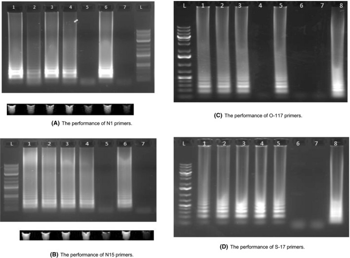
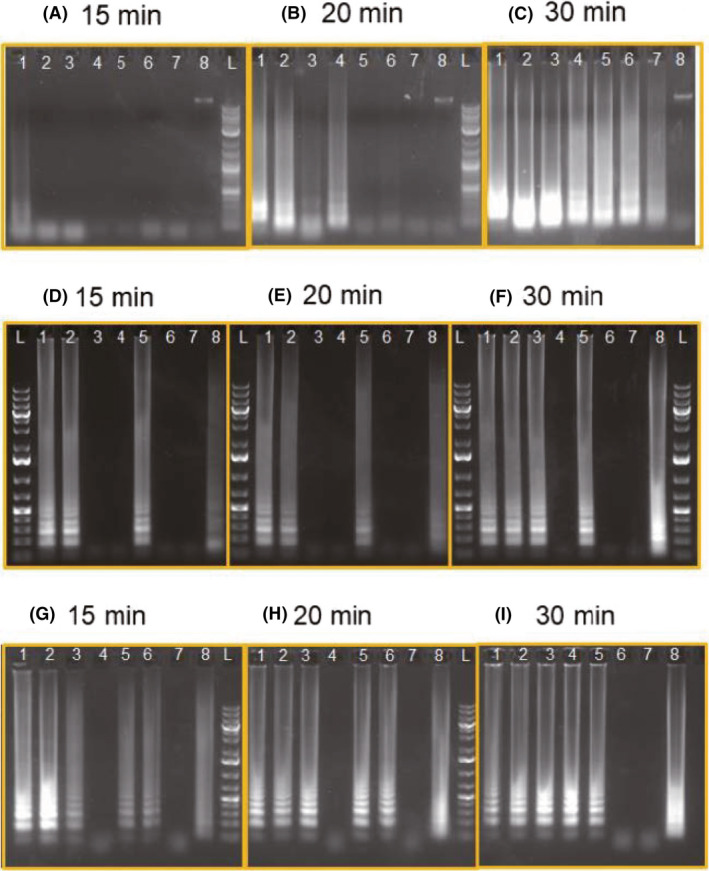
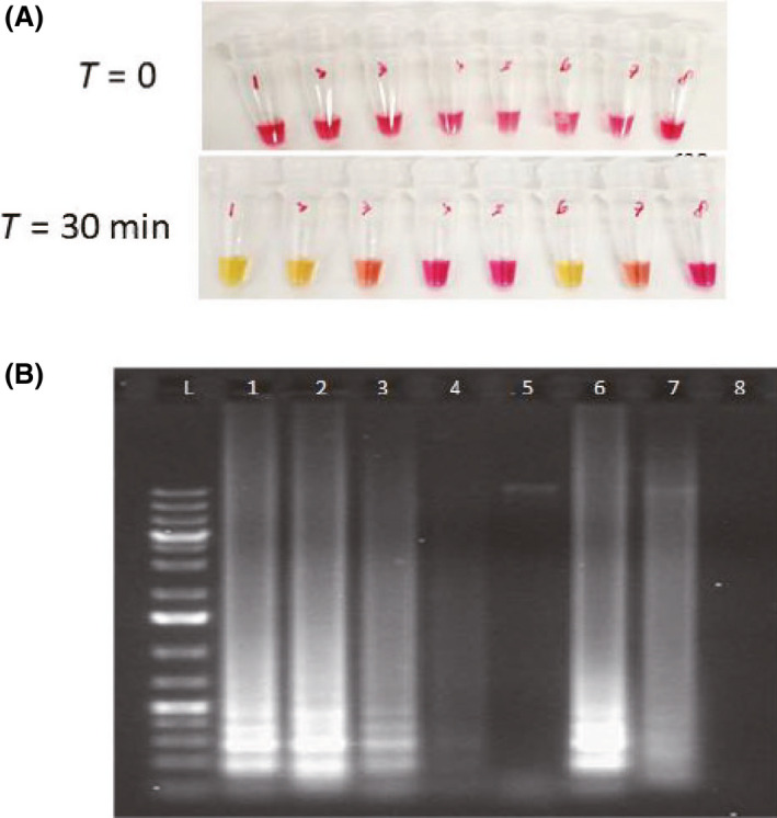
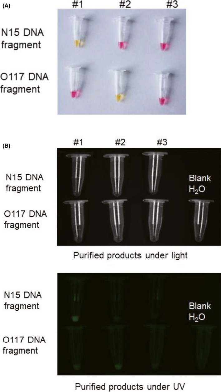
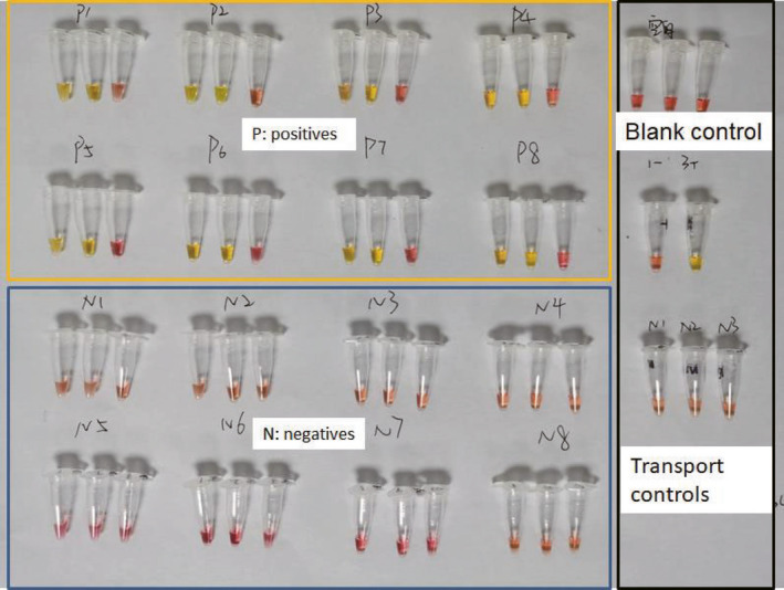
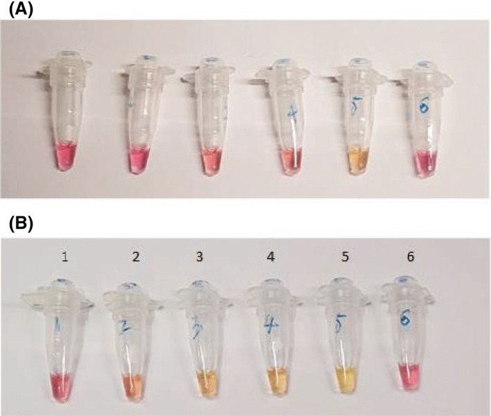
The pandemic coronavirus SARS-CoV-2 in the world has caused a large infected population suffering from COVID-19. To curb the spreading of the virus, WHO urgently demanded an extension of screening and testing; thus, a rapid and simple diagnostic method is needed. We applied a reverse transcription-loop-mediated isothermal amplification (RT-LAMP) to achieve the detection of SARS-CoV-2 in 30 min. We designed four sets of LAMP primers (6 primers in each set), targeting the viral RNA of SARS-CoV-2 in the regions of orf1ab, S gene and N gene. A colorimetric change was used to report the results, which enables the outcome of viral RNA amplification to be read by the naked eye without the need of expensive or dedicated instrument. The sensitivity can be 80 copies of viral RNA per ml in a sample. We validated the RT-LAMP method in a hospital in China, employing 16 clinic samples with 8 positives and 8 negatives. The testing results are consistent with the conventional RT-qPCR. In addition, we also show that one-step process without RNA extraction is feasible to achieve RNA amplification directly from a sample. This rapid, simple and sensitive RT-LAMP method paves a way for a large screening at public domain and hospitals, particularly regional hospitals and medical centres in rural areas.


Conflict of interest statement

None declared.

Figures

Fig. 1
Fig. 1
LAMP results of four different sets of primers targeting the DNA of N, S and Orf1ab gene of SARS‐CoV‐2. A. Performance of N‐1 primer set using N gene DNA sequence. (1) N‐15 primers + N gene DNA (200k copies)*; (2) N15 primers + N gene DNA (200 copies); (3) N15 primers + N gene DNA (200k copies) + Human genome; (4) N15 primers + N gene DNA (200 copies) + Human genome; (5) N15 primers + Human genome; (6) Human β‐actin primers + Human genome; (7) Human β‐actin primers + N gene DNA (200k copies); L, Ladder. LAMP results using fluorescent dye were visualized under UV exposure, which is consistent to the result of gel electrophoresis. B. Performance of N‐15 primer set using N gene DNA sequence. (1) N‐15 primers + N gene DNA (200k copies); (2) N15 primers + N gene DNA (200 copies); (3) N15 primers + N gene DNA (200k copies) + Human genome; (4) N15 primers + N gene DNA (200 copies) + Human genome; (5) N15 primers + Human genome; (6) Human β‐actin primers + Human genome; (7) Human β‐actin primers + N gene DNA (200k copies); L, Ladder. LAMP results using fluorescent dye were visualized under UV exposure, which is consistent to the result of gel electrophoresis. C. Performance of O‐117 primer set using Orf1ab gene DNA sequence. (1) O‐117 primers + Orf1ab gene DNA (200k copies); (2) O‐117 primers + Orf1ab gene DNA (200 copies); (3) O‐117 primers + Orf1ab gene DNA (20 copies); (4) O‐117 primers + Orf1ab gene DNA (2 copies); (5) O‐117 Primers + Orf1ab gene DNA (200 copies) + Human Genome; (6) O‐117 Primers + Human Genome; (7) O‐117 Primers + water (8) Human β‐actin primers + human genomic DNA; (L) Ladder. D. Performance of S‐17 primer set using S gene DNA sequence. (1) S‐17 primers + S gene DNA (200k copies); (2) S‐17 primers + S gene DNA (200 copies); (3) S‐17 primers + S gene DNA (20 copies); (4) S‐17 primers + S gene DNA (2 copies); (5) S‐17 Primers + S gene DNA (200 copies) + Human Genome; (6) S‐17 Primers + Human Genome; (7) S‐17 primers + water (8) Human β‐actin primers + human genomic DNA; (L) Ladder. *200k (200,000) 200, 20 and 2 represent the total copy number of DNA sequence in the reaction mix.
Fig. 2
Fig. 2
Sensitivity and efficiency of RT‐LAMP using N1 and N15 primers (A, B, C). RNA sequence of N gene was derived from in vitro transcription and used as the target. Reaction mixture was sampling at (A) 15 min, (B) 20 min and (C) 30 min. (1) N1 Primers + N gene RNA (200); (2) N1 Primers + N gene RNA (20); (3) N1 Primers + N gene RNA (2); (4) N15 Primers + N gene RNA (200); (5) N15 Primers + N gene RNA (20); (6) N15 Primers + N gene RNA (2); (L) Ladder. Sensitivity and efficiency of RT‐LAMP using O‐117 (D, E, F) and S‐17 primers (G, H, I). RNA of orf1ab and S17 was derived from in vitro transcription and used as the targets. Reaction mixture was sampling at (D, G) 15 min, (E, H) 20 min and (F, I) 30 min. (1) Viral Primers + RNA target (200 k); (2) Viral Primers + RNA target (200); (3) Viral Primers + RNA target (20); (4) Viral Primers + RNA target (2); (5) Viral Primers + RNA target (200) + human genome; (6) Viral Primers + human genome; (7) Viral Primers + Water; (8) Human Primers + human genome
Fig. 3
Fig. 3
Colorimetric RT‐LAMP assay using N15 primers. A. Colour of reaction mixtures before RT‐LAMP. B. Colour of reaction mixtures after RT‐LAMP. C. Gel electrophoresis of reaction mixtures. The intensity of RT‐LAMP products in the gel tallies with the colour of reaction mixtures and provides a semi‐quantitative results. (1) N15 Primers + N gene RNA (200k); (2) N15 Primers + N gene RNA (200); (3) N15 Primers + N gene RNA (20); (4) N15 Primers + N gene RNA (2); (5) N15 Primers + human genome; (6) Human primers + human genome; (7) Human Primers whole human RNA; (8) Colorimetric MasterMix only.
Fig. 4
Fig. 4
The products of RT‐LAMP can be read by both colour change and fluorescent signal, which are consistent. A. Colorimetric display of the RT‐LAMP products. B. Fluorescent image under the UV lamp of the RT‐LAMP products. #1, #2 and #3 samples separately contained N15, O117 and β‐actin primers.
Fig. 5
Fig. 5
Validation of RT‐LAMP using 16 clinical COVID‐19 samples from Shenzhen Luohu People’s hospital. Samples were first tested with conventional RT‐PCR (Table 2), and eight positive samples were labelled ‘P’, and eight negative samples were labelled ‘N’. The components of each test kit are listed in Experimental procedures. Three tubes in each RT‐LAMP assay are #1, #2, #3 in order from left to right. Tube #1 and #2 separately contain O117 and N15 primers targeting the Orf1ab gene and N gene of SARS‐CoV‐2. Tube #3 contain human β‐actin primers. To tube #1 and 2, all positive samples turned yellow, whilst all negative samples remained pink after 30‐minute reaction. The results of RT‐LAMP method are consistent to the results of conventional RT‐PCR (Table 2 and Fig. S1).
Fig. 6
Fig. 6
One‐step process to get the products of nucleic acid amplification. One microlitre of iPSC cells in 0.85% sterile saline was directly added into the LAMP reagents in each tube and incubate for 30 min (A) and 40 min (B). The whole process was 40 min. (1) 0 cells + Human β‐actin primers. (2) 10 cells + Human β‐actin primers. (3) 50 cells + Human β‐actin primers. (4) 100 cells + Human β‐actin primers. (5) Extracted human RNA + Human β‐actin primers. (6) H2O + Human β‐actin primers.

Similar articles

Cited by

References

    1. Boehme, C.C. , Nabeta, P. , Henostroza, G. , Raqib, R. , Rahim, Z. , Gerhardt, M. , et al (2007) Operational feasibility of using loop‐mediated isothermal amplification for diagnosis of pulmonary tuberculosis in microscopy centers of developing countries. J Clin Microbiol 45: 1936–1940. - PMC - PubMed
    1. Cui, J. , Li, F. , and Shi, Z.L. (2019) Origin and evolution of pathogenic coronaviruses. Nat Rev Microbiol 17: 181–192. - PMC - PubMed
    1. Drosten, C. , Gottig, S. , Schilling, S. , Asper, M. , Panning, M. , Schmitz, H. , and Gunther, S. (2002) Rapid detection and quantification of RNA of Ebola and Marburg viruses, Lassa virus, Crimean‐Congo hemorrhagic fever virus, Rift Valley fever virus, Dengue virus, and Yellow fever virus by real‐time reverse transcription‐PCR. J Clin Microbiol 40: 2323–2330. - PMC - PubMed
    1. Espy, M.J. , Uhl, J.R. , Sloan, L.M. , Buckwalter, S.P. , Jones, M.F. , Vetter, E.A. , et al (2006) Real‐time PCR in clinical microbiology: Applications for a routine laboratory testing. Clin Microbiol Rev 19: 165. - PMC - PubMed
    1. Hsieh, K. , Mage, P.L. , Csordas, A.T. , Eisenstein, M. , and Soh, H.T. (2014) Simultaneous elimination of carryover contamination and detection of DNA with uracil‐DNA‐glycosylase‐supplemented loop‐mediated isothermal amplification (UDG‐LAMP). Chem Commun 50: 3747–3749. - PubMed

Publication types

Associated data