. 2020 Apr 25;22(1):91.
doi: 10.1186/s13075-020-02161-8.

Higher interferon score and normal complement levels may identify a distinct clinical subset in children with systemic lupus erythematosus

Affiliations

Higher interferon score and normal complement levels may identify a distinct clinical subset in children with systemic lupus erythematosus

Alessandra Tesser et al. Arthritis Res Ther. .

Abstract

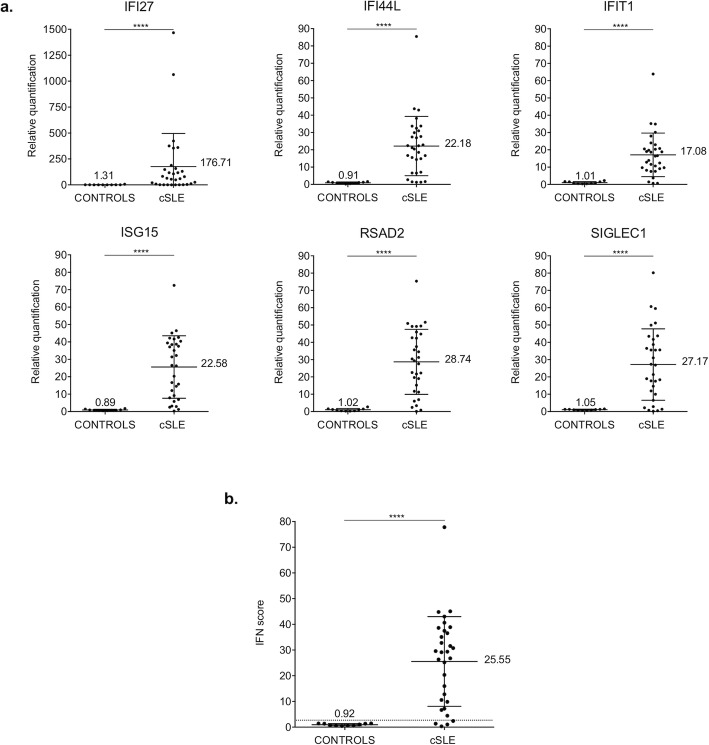
Background: Systemic lupus erythematosus (SLE) is a complex multi-system disease, characterized by both autoimmune and autoinflammatory clinical and laboratory features. The role of type I interferon (IFN) in SLE has been demonstrated from the 2000s, by gene expression analyses showing significant over-expression of genes related to type I IFN signalling pathway (IFN signature). However, several studies questioned the role of measuring the intensity of IFN signature (IFN score) to chase SLE activity. We would assess if the IFN signature can help the clinical and therapeutic stratification of patients with pediatric SLE.

Methods: We measured the IFN score in peripheral whole blood from a series of subjects with childhood-onset SLE and correlated the results with clinical and laboratory parameters.

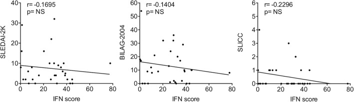
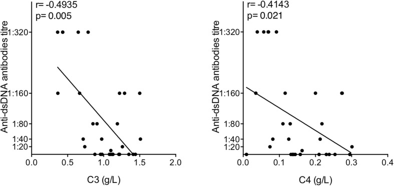
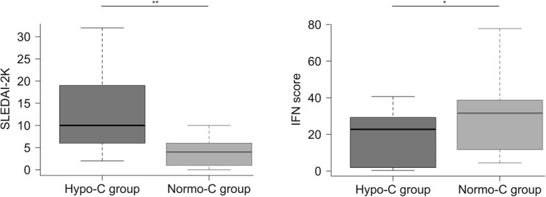
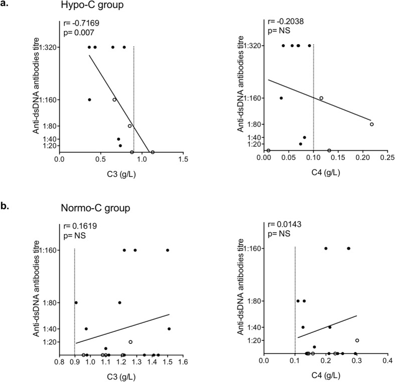
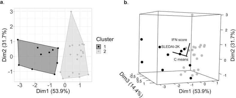
Results: Thirty-one subjects were included in the study, among which the 87% displayed a positive IFN score. The only significant relation was found for high IFN score in subjects with normocomplementemia. No correlation was observed between IFN score and SLEDAI-2K, BILAG-2004 and SLICC. Patients with high IFN score and normal complement levels also presented lower anti-dsDNA antibodies.

Conclusions: The integration between IFN signature analysis and complement levels may easily distinguish two groups of subjects, in which the autoimmune or autoinflammatory component of the disease seems to be prevalent.

Keywords: Autoimmunity; Autoinflammation; Complement; Interferon score; Patients’ stratification; Systemic lupus erythematosus.


Conflict of interest statement

The authors declare that they have no competing interests.

Figures

Fig. 1
Fig. 1
Interferon (IFN) signature analysis in thirty-one patients with childhood-onset systemic lupus erythematosus (cSLE) and ten healthy controls (CONTROLS). a Relative quantification (RQ) of interferon-stimulated genes (ISGs) (IFI27, IFI44L, IFIT1, ISG15, RSAD2, SIGLEC1) normalized on housekeeping genes. b IFN score calculated as the median of the RQ of the ISGs in patients and healthy controls. The dashed line represents the cut off value (2.466) determined by Crow [23, 36], to identify as positive/negative the IFN signature analysis. Results are reported as mean (shown in the figure) ± standard deviation. Data were analyzed with Mann-Whitney non-parametric test (****p < 0.0001)
Fig. 2
Fig. 2
Linear regression analysis between IFN score and SLEDAI-2K, BILAG-2004 and SLICC indices by calculating Spearman’s rank correlation coefficient. p values < 0.05 were considered significant (NS, not significant)
Fig. 3
Fig. 3
Linear regression analysis between anti-dsDNA antibodies and C3 and C4 levels, by calculating Spearman’s rank correlation coefficient. p values < 0.05 were considered significant
Fig. 4
Fig. 4
Comparison between SLEDAI-2K and IFN score values of the hypocomplementemic (Hypo-C group, n = 12) and normocomplementemic (Normo-C group, n = 19) subset. Values distribution is shown by boxplot (Whiskers 5–95 percentile). Data were analyzed with Mann-Whitney non-parametric test (*p < 0.05, **p < 0.01)
Fig. 5
Fig. 5
Linear regression analysis between C3 and C4 levels with anti-dsDNA antibodies in the Hypo-C (a) and Normo-C (b) group, by calculating Spearman’s rank correlation coefficient. p values < 0.05 were considered significant (NS, not significant). The dashed line shows the lower threshold value for C3 (0.9 g/L) and C4 (0.1 g/L). In a, the black dots represent patient with a low level of both C3 and C4, whilst the white dots symbolize subjects with low levels of either C3 or C4; in b, normocomplementemic patients which previously showed low complement levels are indicated by white dots
Fig. 6
Fig. 6
Cluster analysis results (K-means clustering) considering IFN score, SLEDAI-2K and mean complement levels (C means, mean value of C3 and C4 levels normalized on the respective lower threshold) that divide patients into subgroups by similarities. a 2D clusters representation (each dot represents a subject; Dim1 and Dim2 show the higher differences between the main clusters; Cluster 1: Hypo-C group, Cluster 2: Normo-C group). b 3D cluster representation (each sphere represents a patient; Dim3 reports minor difference in the third dimension, accentuated by SLEDAI-2K)

References

    1. Tsokos GC. Systemic lupus erythematosus. N Engl J Med. 2011;365(22):2110–2121. doi: 10.1056/NEJMra1100359. - DOI - PubMed
    1. Mok CC, Lau CS. Pathogenesis of systemic lupus erythematosus. J Clin Pathol. 2003;56(7):481–490. doi: 10.1136/jcp.56.7.481. - DOI - PMC - PubMed
    1. Silva CA, Avcin T, Brunner HI. Taxonomy for systemic lupus erythematosus with onset before adulthood. Arthritis Care Res (Hoboken) 2012;64(12):1787–1793. doi: 10.1002/acr.21757. - DOI - PMC - PubMed
    1. Malattia C, Martini A. Paediatric-onset systemic lupus erythematosus. Best Pract Res Clin Rheumatol. 2013;27(3):351–362. doi: 10.1016/j.berh.2013.07.007. - DOI - PubMed
    1. Tarr T, Dérfalvi B, Győri N, Szántó A, Siminszky Z, Malik A, et al. Similarities and differences between pediatric and adult patients with systemic lupus erythematosus. Lupus. 2015;24(8):796–803. doi: 10.1177/0961203314563817. - DOI - PubMed

Publication types