

Optic chiasm measurements may be useful markers of anterior optic pathway degeneration in neuromyelitis optica spectrum disorders
- PMID: 32335748
- PMCID: PMC7431438
- DOI: 10.1007/s00330-020-06859-w
Optic chiasm measurements may be useful markers of anterior optic pathway degeneration in neuromyelitis optica spectrum disorders
Abstract
Objectives: We aimed to evaluate optic chiasm (OC) measures as potential imaging marker for anterior optic pathway damage assessment in the context of neuromyelitis optica spectrum disorders (NMOSD).
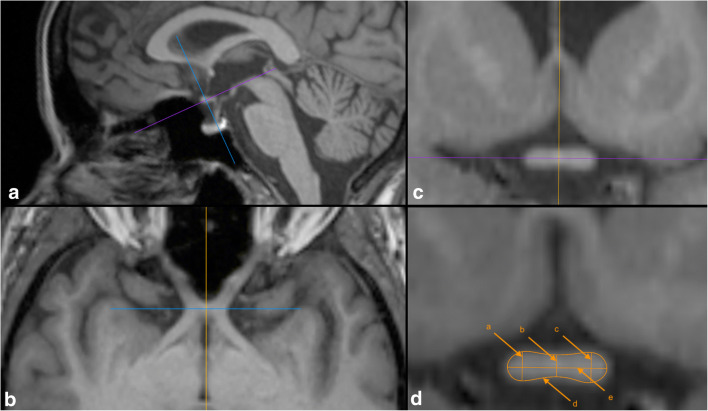
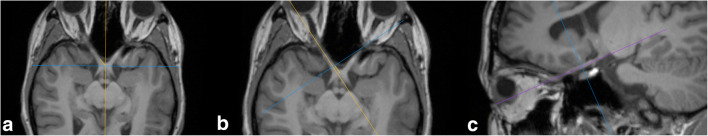
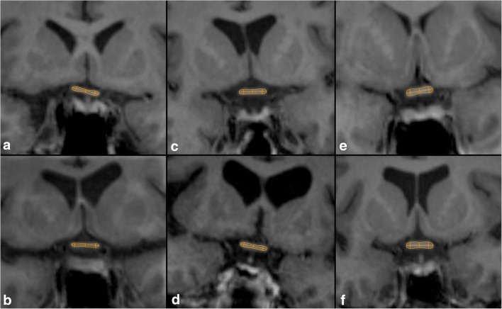
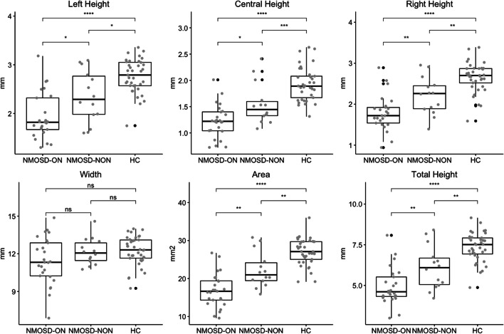
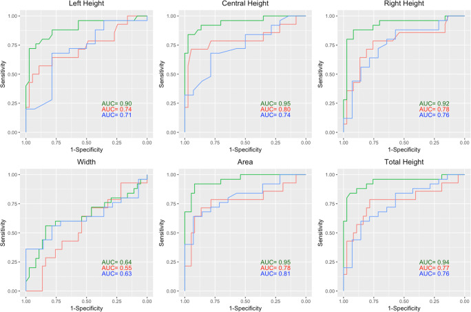
Materials and method: This cross-sectional study included 39 patients exclusively with aquaporin 4-IgG seropositive NMOSD of which 25 patients had a history of optic neuritis (NMOSD-ON) and 37 age- and sex-matched healthy controls (HC). OC heights, width, and area were measured using standard 3D T1-weighted MRI. Sensitivity of these measures to detect neurodegeneration in the anterior optic pathway was assessed in receiver operating characteristics analyses. Correlation coefficients were used to assess associations with structural measures of the anterior optic pathway (optic nerve dimensions, retinal ganglion cell loss) and clinical measures (visual function and disease duration).
Results: OC heights and area were significantly smaller in NMOSD-ON compared to HC (NMOSD-ON vs. HC p < 0.0001). An OC area smaller than 22.5 mm2 yielded a sensitivity of 0.92 and a specificity of 0.92 in separating chiasms of NMOSD-ON from HC. OC area correlated well with structural and clinical measures in NMOSD-ON: optic nerve diameter (r = 0.4, p = 0.047), peripapillary retinal nerve fiber layer thickness (r = 0.59, p = 0.003), global visual acuity (r = - 0.57, p = 0.013), and diseases duration (r = - 0.5, p = 0.012).
Conclusion: Our results suggest that OC measures are promising and easily accessible imaging markers for the assessment of anterior optic pathway damage.
Key points: • Optic chiasm dimensions were smaller in neuromyelitis optica spectrum disorder patients compared to healthy controls. • Optic chiasm dimensions are associated with retinal measures and visual dysfunction. • The optic chiasm might be used as an easily accessible imaging marker of neurodegeneration in the anterior optic pathway with potential functional relevance.
Keywords: Magnetic resonance imaging; Neuromyelitis optica; Optic chiasm; Optic neuritis.
Conflict of interest statement
The authors of this manuscript declare relationships with the following companies:
NS has received travel grants from Biogen Idec and sanofi-aventis/Genzyme.
SA received travel grants from Celgene GmbH, unrelated to this project.
FCO was employed by Nocturne, unrelated to this project.
HZ received research grants from Novartis, unrelated to this project.
KR was supported by the German Ministry of Education and Research (BMBF/KKNMS, Competence Network Multiple Sclerosis) and has received research support from Novartis and Merck Serono as well as speaking fees and travel grants from Guthy Jackson Charitable Foundation, Bayer Healthcare, Biogen Idec, Merck Serono, sanofi-aventis/Genzyme, Teva Pharmaceuticals, Roche, and Novartis.
JBS has received travel grants and speaking fees from Bayer Healthcare, Biogen Idec, Merck Serono, sanofi-aventis/Genzyme, Teva Pharmaceuticals, and Novartis.
FP declares that he has received research grants and speaker’s honoraria from Bayer Healthcare, Teva Pharmaceuticals, Genzyme, Merck and Co., Novartis, and MedImmune. He is also a member of the steering committee for the OCTIMS study (run by Novartis).
AUB is cofounder and shareholder of technology start-ups Motognosis and Nocturne. He is named as inventor on several patent applications describing MS serum biomarkers, perceptive visual computing for motor function assessment and retinal image analysis.
Figures
References
-
- Hickman SJ, Brex PA, Brierley CM, et al. Detection of optic nerve atrophy following a single episode of unilateral optic neuritis by MRI using a fat-saturated short-echo fast FLAIR sequence. Neuroradiology. 2001;43:123–128. - PubMed
-
- Hickman SJ, Toosy AT, Jones SJ, et al. A serial MRI study following optic nerve mean area in acute optic neuritis. Brain. 2004;127:2498–2505. - PubMed
-
- Tian DC, Su L, Fan M, et al. Bidirectional degeneration in the visual pathway in neuromyelitis optica spectrum disorder (NMOSD) Mult Scler. 2018;24:1585–1593. - PubMed
MeSH terms
Substances
LinkOut - more resources
Full Text Sources
Miscellaneous
