Review
. 2020 May;30(2):193-203.
doi: 10.1016/j.nic.2020.01.001. Epub 2020 Mar 27.

Magnetoencephalography Research in Pediatric Autism Spectrum Disorder

Affiliations
Review

Magnetoencephalography Research in Pediatric Autism Spectrum Disorder

Heather L Green et al. Neuroimaging Clin N Am. 2020 May.

Abstract

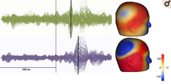
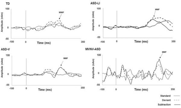
Magnetoencephalography (MEG) research indicates differences in neural brain measures in children with autism spectrum disorder (ASD) compared to typically developing (TD) children. As reviewed here, resting-state MEG exams are of interest as well as MEG paradigms that assess neural function across domains (e.g., auditory, resting state). To date, MEG research has primarily focused on group-level differences. Research is needed to explore whether MEG measures can predict, at the individual level, ASD diagnosis, prognosis (future severity), and response to therapy.

Keywords: ASD; Biomarker; ERD; M100; M50; MEG; MMF.


Figures

Figure 1.
Figure 1.
Representative child with ASD (bottom) shows a delayed M100 response compared to an age-matched typically developing control (top). At left, sensor overlay plots show the auditory evoked response from a typically developing child (green) and a child with ASD (purple). The stimulus onset is marked in gray and the M100 response shown with the black line. At right, M100 magnetic field maps (top: TD; bottom: ASD) From RG Port, AR Anwar, M Ku, et al. Prospective MEG biomarkers in ASD: pre-clinical evidence and clinical promise of electrophysiological signatures. Yale J Biol Med 2015. Mar 4;88(1): p.27; with permission.
Figure 2.
Figure 2.
Source response waveforms from superior temporal gyrus. Vertical lines indicate stimulus onset (0 ms). Arrows indicate MMF latency in a representative TD child (182 ms), verbal ASD without language impairment (214 ms), ASD and language impairment (222 ms), and ASD MVNV (270 ms). The solid gray line indicates the standard response, the dashed gray line indicates the deviant response, and the solid black line their subtraction to yield the difference wave. From Matsuzaki, J., E. S. Kuschner, L. Blaskey, et al. Abnormal auditory mismatch fields are associated with communication impairment in both verbal and minimally verbal/nonverbal children who have autism spectrum disorder. Autism Res 2019. Aug;12(8):1225–1235; with permission.
Figure 3.
Figure 3.
Scatterplot of Clinical Evaluation of Language-Fourth Edition (CELF-4), Core language index scores (x-axis) and right STG pre-stimulus gamma activity (y-axis, 30–50 Hz) in individuals with ASD. In ASD lower CELF-4 scores were associated with increased right-hemisphere pre-stimulus gamma activity. From Edgar, J. C., S. Y. Khan, L. Blaskey, et al. Neuromagnetic oscillations predict evoked-response latency delays and core language deficits in autism spectrum disorders. J Autism Dev Disord 2015; 45(2):395–405; with permission.
Figure 4.
Figure 4.
(a) Sensory waveforms grand-averaged across subject and token, time-locked to token onset. Shaded area (red) indicates the time window used in ERD measurements. (b) Grand average spectro-temporal plots (TFR’s) for the left and right auditory cortex source locations. From L Bloy, K Shwayder, L Blaskey, et al. A Spectrotemporal Correlate of Language Impairment in Autism Spectrum Disorder. J Autism Dev Disord 2019; Aug 49(8): 3181–3190; with permission.
Figure 5.
Figure 5.
Scatterplots showing associations between age (x axis) and peak alpha frequency (y axis) for TDC (blue) and ASD (red). As detailed in Section 3, a significant interaction term indicated group slope differences. ***p < 0.001. From JC Edgar, M Dipiero, E McBride. Abnormal maturation of the resting-state peak alpha frequency in children with autism spectrum disorder. Hum Brain Mapp 2019. Aug 1;40(11):3288–3298; with permission.
Figure 6.
Figure 6.
(a) Scatterplots showing associations between nonverbal IQ (x axis) and peak alpha frequency (y axis) for TDC (blue) and ASD (red). (b) Nonverbal IQ and peak alpha frequency associations shown for the younger (<10-years-old; left plot) and older children (>10-years-old; right plot). *p < 0.05. From JC Edgar, M Dipiero, E McBride. Abnormal maturation of the resting-state peak alpha frequency in children with autism spectrum disorder. Hum Brain Mapp 2019. Aug 1;40(11):3288–3298; with permission.

Similar articles

Cited by

References

    1. Aaltonen O, Niemi P, Nyrke T & Tuhkanen M (1987) Event-related brain potentials and the perception of a phonetic continuum. Biol Psychol, 24, 197–207. - PubMed
    1. Alho K (1995) Cerebral generators of mismatch negativity (MMN) and its magnetic counterpart (MMNm) elicited by sound changes. Ear Hear, 16, 38–51. - PubMed
    1. Alvarez Amador A, Sosa P. A. Valdés, Marqui R. D. Pascual, Garcia L. Galan, Lirio R. Biscay & Bayard J. Bosch (1989) On the structure of EEG development. Electroencephalogr Clin Neurophysiol, 73, 10–9. - PubMed
    1. American Psychiatric Association [APA], D.-T. F. 2013. Diagnostic and statistical manual of mental disorders: DSM-5 (5th Ed.). Arlington, VA: American Psychiatric Publishing, Inc.
    1. Baio J, Wiggins L, Chistenson DL, Maenner MJ, Daniels J, Warren Z, Kurzius-Spencer M, Zahorodny W, Rosenberg CR, White T, Durkin M, Imm P, Nikolau L, Yeargin-Allsopp M, Lee LC, Harrington R, Lopez M, Fitzgerald RT, Hewitt A, Pettygrove S, Constantino JN, Vehorn A, Shenouda J, Hall-Lande J, Van Naarden Braun K & Dowling N (2018) Prevalence of autism spectrum disorder among children aged 8 years - autism and developmental disabilities monitoring network, 11 sites, United States,2014. Centers for Disease Control and Prevention: Morbidity and Mortality Weekly Report, 67, 1–23. - PMC - PubMed