. 2021 Mar 1;106(3):692-700.
doi: 10.3324/haematol.2019.236513.

Stem cell factor is implicated in microenvironmental interactions and cellular dynamics of chronic lymphocytic leukemia

Affiliations

Stem cell factor is implicated in microenvironmental interactions and cellular dynamics of chronic lymphocytic leukemia

George I Gavriilidis et al. Haematologica. .

Abstract

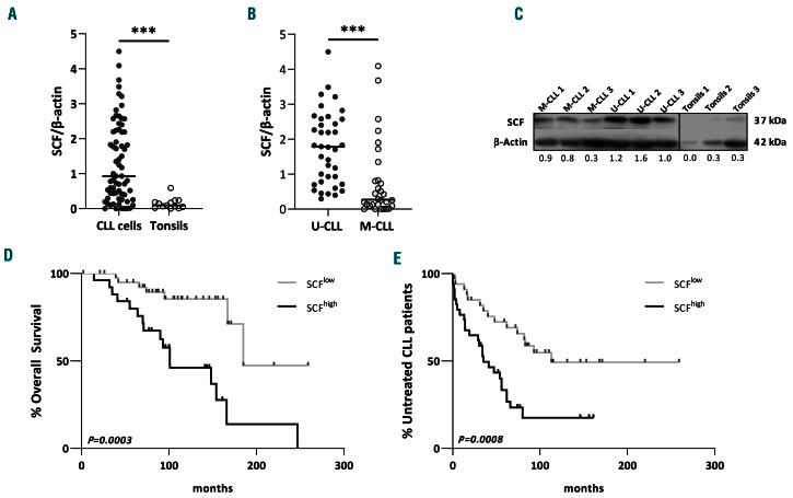
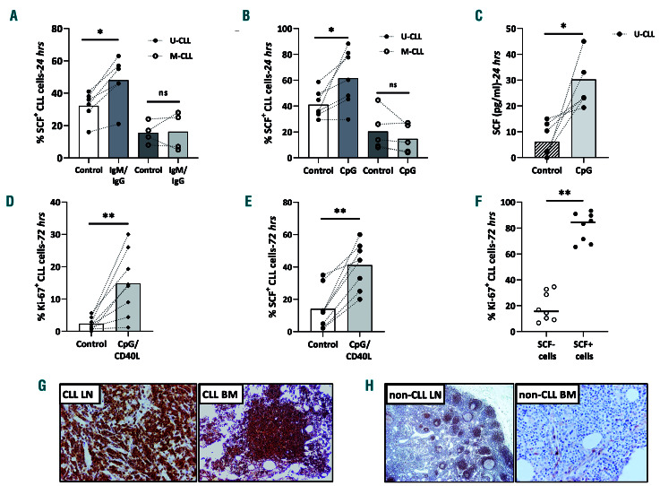
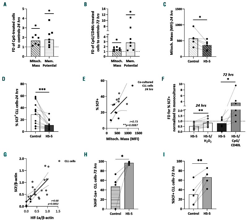
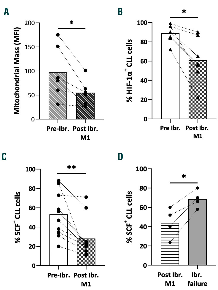
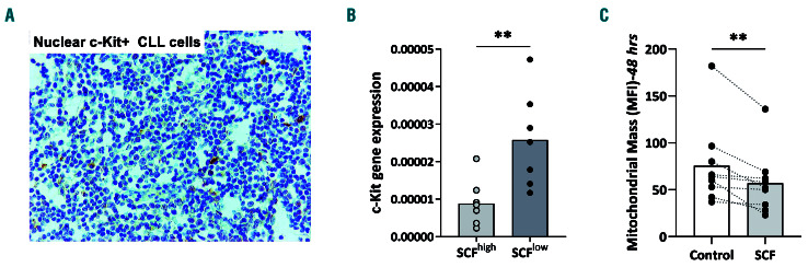
The inflammatory cytokine stem cell factor (SCF, ligand of c-kit receptor) has been implicated as a pro-oncogenic driver and an adverse prognosticator in several human cancers. Increased SCF levels have recently been reported in a small series of patients with chronic lymphocytic leukemia (CLL), however its precise role in CLL pathophysiology remains elusive. In this study, CLL cells were found to express predominantly the membrane isoform of SCF, which is known to elicit a more robust activation of the c-kit receptor. SCF was significantly overexpressed in CLL cells compared to healthy tonsillar B cells and it correlated with adverse prognostic biomarkers, shorter time-to-first treatment and shorter overall survival. Activation of immune receptors and long-term cell-cell interactions with the mesenchymal stroma led to an elevation of SCF primarily in CLL cases with an adverse prognosis. Contrariwise, suppression of oxidative stress and the BTK inhibitor ibrutinib lowered SCF levels. Interestingly, SCF significantly correlated with mitochondrial dynamics and hypoxia-inducible factor-1a which have previously been linked with clinical aggressiveness in CLL. SCF was able to elicit direct biological effects in CLL cells, affecting redox homeostasis and cell proliferation. Overall, the aberrantly expressed SCF in CLL cells emerges as a key response regulator to microenvironmental stimuli while correlating with poor prognosis. On these grounds, specific targeting of this inflammatory molecule could serve as a novel therapeutic approach in CLL.


Figures

Figure 1.
Figure 1.
Stem cell factor is overexpressed in chronic lymphocytic leukemia cells and correlated with adverse prognosis. (A, B) Comparison of the expression of stem cell factor (SCF) protein in chronic lymphocytic leukemia (CLL) cells compared to healthy tonsillar B cells (A) and in IG-unmutated (U-CLL) compared to IG-mutated (M-CLL) cells, showing overexpression in U-CLL cases (B). The dot plots show SCF protein levels normalized to β-actin from western blot studies. Each dot represents one case. (C) Representative western blot for the distinct SCF probing in samples from healthy tonsils, U-CLL and M-CLL cases. (D, E) SCFhigh cases have shorter overall survival (OS) (D) and shorter time-to-first treatment (TTFT) (E) compared to SCFlow cases, as shown by Kaplan-Meier OS and TTFT curves for CLL patients with high SCF expression (normalized SCF protein levels to β-actin >1.7 for OS and >0.93 for TTFT) and low SCF expression (normalized SCF protein expression to β-actin ≤1.7 for OS and ≤0.93 for TTFT). Cut-off points were generated using time-dependent receiver operating characteristic curve analysis. The Mann–Whitney U and the log-rank test were applied as appropriate to evaluate statistical significance; *P<0.05, **P<0.01, ***P<0.001.
Figure 2.
Figure 2.
Microenvironmental triggering induces stem cell factor in chronic lymphocytic leukemia cells. (A) Comparison of stem cell factor (SCF)+ cells (flow cytometry for viable/SCF+ chronic lymphocytic leukemia [CLL] cells) from IG-unmutated CLL (U-CLL) and IG-mutated (M-CLL) cases, untreated (Control) cases and cases treated with IgM/IgG. (B) Comparison of SCF+ cells (flow cytometry for viable/SCF+ CLL cells) from U-CLL and M-CLL cases, untreated (Control) cases and cases treated with CpG. (C) Comparison of secreted SCF (pg/mL; determined by enzyme-linked immunosorbent assay in cellular supernatants) from U-CLL cells, untreated (Control) and CpG-treated cells. (D, E) Comparison of proliferating cells (Ki-67+) (D) and SCF+ cells (%) (E) in untreated (Control) and CpG/CD40L-treated CLL cases, as determined by flow cytometry. (F) The majority of proliferating (Ki-67+) CLL cells were also SCF+ as determined by flow cytometry. (G) Lymph node (left) and the corresponding bone marrow biopsy (right) from the same CLL patient showing CLL invading cells with aberrant SCF expression. (H) Reactive, non-CLL lymph node (left) exhibiting SCF positivity mostly in mantle zones but not in germinal centers and reactive, non-CLL bone marrow (right) exhibiting scant SCF positivity in scarce immune and endothelial cells. Interconnected dots represent one case in two different conditions while bars represent median values. The Wilcoxon P test was applied to evaluate statistical significance; *P<0.05, **P<0.01, ***P<0.001. FD: fold difference.
Figure 3.
Figure 3.
Mitochondrial redox homeostasis modulates stem cell factor in chronic lymphocytic leukemia. (A) Mitochondrial mass (determined by mean fluorescent intensity, MFI) and membrane potential (% +) of CpG-treated IG-unmutated chronic lymphocytic leukemia (U-CLL) cells: flow cytometry analysis of viable, CpG-treated U-CLL cells normalized to respective untreated controls. (B) Mitochondrial mass (MFI) and membrane potential (% +) of U-CLL cells treated with CpG/CD40L: flow cytometry analysis of viable CpG/CD40L-treated U-CLL cells normalized to respective untreated controls. (C) Mitochondrial mass (MFI) of CLL cells co-cultured with the mesenchymal HS-5 cells. Interconnected dots represent one case in two different conditions and bars represent median values. (D) Stem cell factor (SCF) protein expression in CLL cells from short-term co-cultures with HS-5 cells: flow cytometry analysis for viable CD19+/SCF+ CLL cells. Interconnected dots represent one case in two different conditions while bars represent median values. (E) Spearman correlation of SCF protein expression and mitochondrial mass in CLL cells cultured alone (gray dots) and co-cultured with HS-5 cells (black dots). The x axis represents the MFI of MitoGreen probing for mitochondrial mass while the y axis the percentage viable CD19+/SCF+ CLL cells. (F) SCF protein expression in CLL cells co-cultured with HS-5 cells ± H2O2 and ± CpG/CD40: flow cytometry analysis of viable CD19+/SCF+ co-cultured CLL cells normalized to viable CD19+/SCF+ monocultured CLL cells. (G) Spearman correlation of SCF protein expression and HIF-1α protein expression in healthy tonsillar B cells (black dots) and CLL B cells (gray dots). The x axis represents HIF-1a protein levels while the y axis represents SCF protein levels, both normalized to β-actin; data are derived from western blot studies. (H, I) Protein expression of hypoxia-inducible factor-1α (HIF-1a) (H) and SCF (I) in SCFhigh U-CLL cells from long-term co-cultures (72 h) with HS-5 cells: flow cytometry analysis for viable CD19+/SCF+ CLL cells. Interconnected dots represent one case in two different conditions while bars represent median values. The Wilcoxon P test was applied to evaluate statistical significance. *P<0.05, **P<0.01, ***P<0.001. FD: fold difference.
Figure 4.
Figure 4.
Ibrutinib leads to downregulation of mitochondrial mass, hypoxiainducible factor-1a and stem cell factor. (A) Mitochondrial mass in chronic lymphocytic leukemia (CLL) samples before and after 1 month of ibrutinib therapy determined by flow cytometry analysis for viable CLL cells. (B) Hypoxia-inducible factor (HIF)-1a protein expression in CLL samples before and after 1 month of ibrutinib therapy determined by flow cytometry analysis for viable HIF-1a+ CLL cells. (C) Stem cell factor (SCF) protein expression in CLL samples before and after 1 month of ibrutinib therapy determined by flow cytometry for viable SCF+ CLL cells. (D) SCF protein expression (determined by flow cytometry for viable SCF+ CLL cells) in samples from CLL patients after 1 month of ibrutinib therapy and loss of response to treatmment. Interconnected dots represent one case in two different conditions and bars represent median values. The Wilcoxon P test was applied to evaluate statistical significance. *P<0.05, **P<0.01. MFI: mean fluorescence intensity; ibr: ibrutinib
Figure 5.
Figure 5.
Functional implications of stem cell factor in chronic lymphocytic leukemia cells. (A) Immunohistochemical staining showing weak nuclear immunopositivity for c-Kit of the scarce chronic lymphocytic leukemia (CLL) cells residing in the lymph nodes. (B) Real-time quantitative polymerase chain reaction analysis of KIT transcripts in stem cell factor (SCF)high CLL cases and SCFlow CLL cases. (C) Mitochondrial mass in CLL cells treated or not (control) with 100 ng/mL SCF for 48 h. Interconnected dots represent one case in two different conditions and bars represent median values. The Wilcoxon P test was applied to evaluate statistical significance; *P<0.05, **P<0.01, ***P<0.001. FD: fold difference.

Similar articles

Cited by

References

    1. Rozovski U, Keating MJ, Estrov Z. Targeting inflammatory pathways in chronic lymphocytic leukemia. Crit Rev Oncol Hematol. 2013;88(3):655-666. - PMC - PubMed
    1. Fabbri G, Dalla-Favera R. The molecular pathogenesis of chronic lymphocytic leukaemia. Nat Rev Cancer. 2016;16(3):145-162. - PubMed
    1. Schulz A, Toedt G, Zenz T, Stilgenbauer S, Lichter P, Seiffert M. Inflammatory cytokines and signaling pathways are associated with survival of primary chronic lymphocytic leukemia cells in vitro: a dominant role of CCL2. Haematologica. 2011;96(3):408-416. - PMC - PubMed
    1. Farinello D, Wozińska M, Lenti E, et al. . A retinoic acid-dependent stroma-leukemia crosstalk promotes chronic lymphocytic leukemia progression. Nat Commun. 2018;9(1):1787. - PMC - PubMed
    1. Yan X-J, Dozmorov I, Li W, et al. . Identification of outcome-correlated cytokine clusters in chronic lymphocytic leukemia. Blood. 2011;118(19):5201-5210. - PMC - PubMed

Publication types