. 2020 Jun;122(12):1825-1836.
doi: 10.1038/s41416-020-0836-4. Epub 2020 Apr 27.

LncRNA-HNF1A-AS1 functions as a competing endogenous RNA to activate PI3K/AKT signalling pathway by sponging miR-30b-3p in gastric cancer

Affiliations

LncRNA-HNF1A-AS1 functions as a competing endogenous RNA to activate PI3K/AKT signalling pathway by sponging miR-30b-3p in gastric cancer

Hai-Ting Liu et al. Br J Cancer. 2020 Jun.

Abstract

Background: Accumulating evidence demonstrated that long noncoding RNAs (lncRNAs) played important regulatory roles in many cancer types. However, the role of lncRNAs in gastric cancer (GC) progression remains unclear.

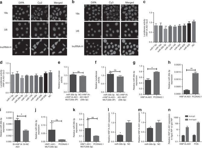
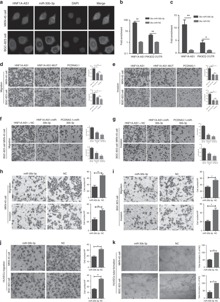
Methods: RT-qPCR assay was performed to detect the expression of HNF1A-AS1 in gastric cancer tissues and the non-tumourous gastric mucosa. Overexpression and RNA interference approaches were used to investigate the effects of HNF1A-AS1 on GC cells. Insight into competitive endogenous RNA (ceRNA) mechanisms was gained via bioinformatics analysis, luciferase assays and an RNA-binding protein immunoprecipitation (RIP) assay, RNA-FISH co-localisation analysis combined with microRNA (miRNA)-pulldown assay.

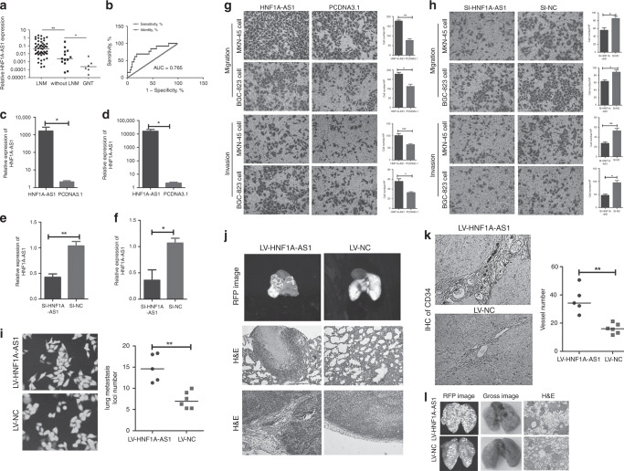
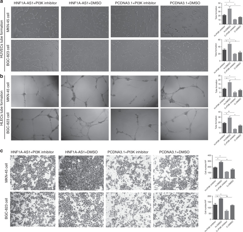
Results: This study displayed that revealed expression of HNF1A-AS1 was associated with positive lymph node metastasis in GC. Moreover, HNF1A-AS1 significantly promoted gastric cancer invasion, metastasis, angiogenesis and lymphangiogenesis in vitro and in vivo. In addition, HNF1A-AS1 was demonstrated to function as a ceRNA for miR-30b-3p. HNF1A-AS1 abolished the function of the miRNA-30b-3p and resulted in the derepression of its target, PIK3CD, which is a core oncogene involved in the progression of GC.

Conclusion: This study demonstrated that HNF1A-AS1 worked as a ceRNA and promoted PI3K/AKT signalling pathway-mediated GC metastasis by sponging miR-30b-3p, offering novel insights of the metastasis mechanism in GC.


Conflict of interest statement

The authors declare no competing interests.

Figures

Fig. 1
Fig. 1. HNF1A-AS1 promotes the migration, invasion and metastasis abilities of GC cells in vivo and in vitro.
a HNF1A-AS1 expression was demonstrated by RT-qPCR assays in human gastric cancer tissues with lymph node metastasis (LNM), without lymph node metastasis (without LNM) and non-tumour gastric mucosa. b Receiver- operating characteristic (ROC) curve showed that HNF1A-AS1 expression could effectively distinguish the cases with LNM from those without LNM in patients with gastric cancers. cf The overexpression (c, d) and knockdown (e, f) efficiency of HNF1A-AS1 in both GC cells. g Transwell assays demonstrated that HNF1A-AS1 enhanced cell migration and invasion abilities in MKN-45 and BGC-823 cells (×200). Three independent experiments were performed, and data are presented as mean ± SD. h Transwell assays indicated that HNF1A-AS1 knockdown inhibited cell migration and invasion abilities in MKN-45 and BGC-823 cells (×200). Three independent experiments were performed, and data are presented as mean ± SD. i HNF1A-AS1 was overexpressed stably in MKN-45 cells, with GFP as marker gene (left, ×200). The number of lung metastatic foci in the LV5-HNF1A-AS1 group was more than that in the LV5-NC group (right). j In total, 1.5 × 106 MKN-45 cells transfected with LV5-HNF1A-AS1 vector or LV5-NC vector were injected into the axillary fossa of the mice. Compared with the LV5-NC group, more stroma, muscle invasion or lung metastasis was observed in the LV5-HNF1A-AS1 group. Representative lung metastasis locus was shown in IVIS system imaging (top) and H&E staining (middle, ×100). Representative stromal invasion was shown in H&E staining (bottom, ×100). k IHC assays of CD-34 showed that the blood vessel density in LV5-HNF1A-AS1 group remarkably increased than that in the LV-NC group (×100). l In total, 2.5 × 106 MKN-45 cells transfected with LV5-HNF1A-AS1 vector or LV5-NC vector were injected into the tail vein of the mice. The number of lung metastatic foci in the LV5-HNF1A-AS1 group was more than that in the LV5-NC group. Representative lung metastasis loci were shown in IVIS system imaging (left), bright-field gross imaging (middle, ×100) and H&E staining (right, ×100), with the quantifiable results shown in Fig. 1i (right). *P < 0.05, **P < 0.01, ***P < 0.001.
Fig. 2
Fig. 2. HNF1A-AS1 promotes HUVEC angiogenesis, HLEC lymphangiogenesis and regulates PIK3CD, PIK3R1 and AKT3 expression.
ad Cell culture supernatants of MKN-45 and BGC-823 cells transiently transfected with the HNF1A-AS1 or the negative control were collected. The cell culture supernatant from the HNF1A-AS1-transfected groups rather than the negative control groups promoted the migration (a, ×200), proliferation (b, ×40) and tube formation (c, ×40) capabilities of HUVECs and also enhanced tube- formation abilities of HLECs (d, ×40). Three independent experiments were performed, and data are presented as mean ± SD. e, f RT-qPCR assays showed that HNF1A-AS1 overexpression increased PIK3CD, PIK3R1 and AKT3 mRNA level in MKN-45 cells (e) and BGC-823 cells (f). Three independent experiments were performed, and data are presented as mean ± SD. g Western blotting assays demonstrated that HNF1A-AS1 overexpression enhanced PIK3CD, PIK3R1 and AKT3 protein expression. Three independent experiments were performed, and data are presented as mean ± SD. h Western blotting assays demonstrated that knockdown of HNF1A-AS1 by siRNA decreased PIK3CD, PIK3R1 and AKT3 protein expression. Three independent experiments were performed, and data are presented as mean ± SD. I, j The secretion level of VEGF-A (i) and VEGF-C (j) significantly enhanced in the culture supernatant from the HNF1A-AS1-transfected MKN-45 cells, compared with the negative control groups, detected by ELISA. Three independent experiments were performed, and data are presented as mean ± SD. k, l The GC cell line MKN-45 cells were transfected with pcDNA3.1 or pcDNA3.1-HNF1A-AS1, and the PI3K/AKT inhibitor NVP-BKM-120 (1 μM) was then added; DMSO was added as a control. In pcDNA3.1-HNF1A-AS1-transfected GC cells, VEGF-A (k) and VEGF-C (l) protein levels decreased significantly with NVP-BKM-120 treatment compared with those of DMSO-treated GC cells. *P < 0.05, **P < 0.01, ***P < 0.001.
Fig. 3
Fig. 3. The effect of PI3K inhibition on HNF1A-AS1-mediated biological functions.
ac The GC cell line MKN-45 cells were transfected with pcDNA3.1 or pcDNA3.1-HNF1A-AS1, and the PI3K/AKT inhibitor NVP-BKM-120 was then added; DMSO was added as a control. NVP-BKM120 (PI3K inhibitor) treatment rescued the tube formation of HUVECs (a, ×40) and HLECs (b, ×40), and migration abilities (c, ×100) of GCs enhanced by HNF1A-AS1. *P < 0.05, **P < 0.01, ***P < 0.001.
Fig. 4
Fig. 4. HNF1A-AS1 functions as a ceRNA with miR-30b-3p.
a, b RNA fluorescent in situ hybridisation of the LncRNA-HNF1A-AS1 in MKN-45 cells (a) and BGC-823 cells (b). The 18 s and U6 probe were respectively used as a cytoplasm and nucleus control (×200). c, d Luciferase assays showed that miR-30b-3p decreased luciferase activity of pmirGLO-HNF1A-AS1 in MKN-45 (c) and BGC-823 (d) cells. e, f Luciferase assays indicated that miR-30b-3p did not decrease luciferase activity of MUT-1 in MKN-45 cells (e) and BGC-823 cells (f). g–k The expression level of miR-30b-3p was detected by RT-qPCR following HNF1A-AS1 overexpression (g, h), knockdown (i) and HNF1A-AS1-MUT-30b-3p (j, k) overexpression in GC cells. l, m RT-qPCR assays showed that miR-30b-3p decreases the expression level of HNF1A-AS1 in MKN-45 cells (l) and BGC-823 cells (m). n HNF1A-AS1 was enriched in Ago2 immunoprecipitates relative to control IgG immunoprecipitates from BGC-823 cell extracts. All the above experiments were performed in three independent experiments, and data are presented as mean ± SD. *P < 0.05, **P < 0.01, ***P < 0.001.
Fig. 5
Fig. 5. MiR-30b-3p abolished the HNF1A-AS1-mediated biological effects.
a Co-localisation between HNF1A-AS1 and miR-30b-3p was observed by RNA-FISH in MKN-45 and BGC-823 cells; FAM-conjugated miR-30b-3p probes and Cy3-labelled HNF1A-AS1 probes were used for staining, followed by counterstain with DAPI (×200). b, c RT-qPCR analysis of HNF1A-AS1 or PIK3CD 3′-UTR levels in the streptavidin-captured fractions from MKN-45 cells (b) and BGC-823 cell (c) lysates after transfection with biotinylated miR-30b-3p or control NC. d, e Transwell assays demonstrated that HNF1A-AS1 with miR-30b-3p-mutated binding sites exerted a weaker effect on GC migration and invasion abilities compared with wild-type HNF1A-AS1 in MKN-45 cells (d) and BGC-823 cells (e) (×100). Three independent experiments were performed, and data are presented as mean ± SD. f, g The migration (f) and invasion (g) abilities were enhanced in HNF1A-AS1-overexpression group, while miR-30b-3p mimics reversed the promotion of migration and invasion capabilities by HNF1A-AS1 (×200). Three independent experiments were performed, and data are presented as mean ± SD. h, i MiR-30b-3p significantly inhibited migration and invasion abilities in MKN-45 (h) and BGC-823 cells (i) (×100). j Migration abilities of HUVECs were dramatically inhibited in both GC cells and transfected groups (×200). k MiR-30b-3p significantly inhibited tube-formation abilities of HUVECs in both GC cells and transfected groups (×40). All the above experiments were performed in three independent experiments, and data are presented as mean ± SD. *P < 0.05, **P < 0.01, ***P < 0.001.
Fig. 6
Fig. 6. MiR-30b-3p exerts tumour-suppressive functions through regulation of PI3K/AKT signalling in GC.
a The schematic of miR-30b-3p in PIK3CD 3′-UTR or mutant sequences. be MiR-30b-3p could reduce the luciferase activities in MKN-45 cells (b) and BGC-823 cells (c) transfected with the PIK3CD 3′-UTR-WT vector, while it had no effect on the PIK3CD 3′-UTR-MUT vector MKN-45 cells (d) and BGC-823 cells (e). f MiR-30b-3p suppressed the PIK3CD and AKT3 protein expression in MKN-45 and BGC-823 cells. g, h Luciferase activity assay showed that miR-30b-3p did not induce a significant decrease in luciferase activity of pmirGLO-AKT3, indicating that AKT3 is not a direct target gene of miR-30b-3p in MKN-45 (g) and BGC-823 cells (h). I, j Luciferase activity assay indicated that overexpression of HNF1A-AS1, but not the HNF1A-AS1 (miR-30b-mut), increased the luciferase activity of pmirGLO-PIK3CD, while ectopic expression of miR-30b-3p diminished this upregulation in MKN-45 (i) and BGC-823 cells (j). k, l RT-qPCR analysis of HNF1A-AS1 or PIK3CD 3′-UTR levels in the streptavidin-captured fractions from MKN-45 cells (k) and BGC-823 cell (l) lysates after transfection with biotinylated miR-30b-3p and pcDNA3.1-HNF1A-AS1 or control group. m, n RT-qPCR analysis of HNF1A-AS1 or PIK3CD 3′-UTR levels in the streptavidin-captured fractions from MKN-45 cells (m) and BGC-823 cell (n) lysates after transfection with biotinylated miR-30b-3p and pcDNA3.1-PIK3CD 3′-UTR or control group. o, p The expression of miR-30b-3p (o) was decreased, while the protein expression of PIK3CD and AKT3 (p) was increased in xenograft tumour tissues of the LV5-HNF1A-AS1 group, compared with those of the LV-NC group. q Graphic model as discussed in the text. lncRNA-HNF1A-AS1 acts as a ceRNA, activates PI3K/AKT signalling by competitively binding to miR-30b-3p, which exhibits oncogenic properties in GC. All the above experiments were performed in three independent experiments, and data are presented as mean ± SD. *P < 0.05, **P < 0.01, ***P < 0.001.

Similar articles

Cited by

References

    1. Hartgrink HH, Jansen EP, van Grieken NC, van de Velde CJ. Gastric cancer. Lancet. 2009;374:477–490. - PMC - PubMed
    1. Shi Y, Zhou Y. The role of surgery in the treatment of gastric cancer. J. surgical Oncol. 2010;101:687–692. - PubMed
    1. Duraes C, Almeida GM, Seruca R, Oliveira C, Carneiro F. Biomarkers for gastric cancer: prognostic, predictive or targets of therapy? Virchows Arch.: Int. J. Pathol. 2014;464:367–378. - PubMed
    1. Guo G, Kang Q, Zhu X, Chen Q, Wang X, Chen Y, et al. A long noncoding RNA critically regulates Bcr-Abl-mediated cellular transformation by acting as a competitive endogenous RNA. Oncogene. 2015;34:1768–1779. - PubMed
    1. Sun M, Nie F, Wang Y, Zhang Z, Hou J, He D, et al. LncRNA HOXA11-AS promotes proliferation and invasion of gastric cancer by scaffolding the chromatin modification factors PRC2, LSD1, and DNMT1. Cancer Res. 2016;76:6299–6310. - PubMed

Publication types

MeSH terms